

Do NOT confuse Alpha Thalessemia with Beta Thelassemia. Even tho Thalassemia minor is MOST similar to Alpha trait, the number of alleles affected is different in each condition.
If you have too little Alpha to make your rbc, you'll pack it with lots of Beta - which is abnormal.
Hg H disease bc in electrophoresis that's the most H, not A as in normal Hgb electrophoresis.
Usu. looking at lab with normal rbc but microcytic, can kinda make out that it's Thalessemia, electrophoresis serves little if any purpose other than genetic counseling later on.
 Most commonly seen in adult clinicals: Trait Alpha thalassemia
Hg H & Alpha Thalassemia Major: usu. already dx in peds or neonatology or Maternal Fetal Medicine
Trait: more problems if too heavy menstruation.
Alpha Major: usu. very bad, already
History: yeah I was told I had some type of anemia but I've never had any symptoms.
Lab: usu. normal RBC, low Hgb, Low MCV (microcytic anemia) however rbc is normal bc the body is able to make enuf rbc with the Hgb & iron on hands. Whereas iron deficiency anemia, the body is unable to make enuf rbc due to not even having enuf iron to even make it.
~by Qt
If you have too little Alpha to make your rbc, you'll pack it with lots of Beta - which is abnormal.
Hg H disease bc in electrophoresis that's the most H, not A as in normal Hgb electrophoresis.
Usu. looking at lab with normal rbc but microcytic, can kinda make out that it's Thalessemia, electrophoresis serves little if any purpose other than genetic counseling later on.
Hg H & Alpha Thalassemia Major: usu. already dx in peds or neonatology or Maternal Fetal Medicine
Trait: more problems if too heavy menstruation.
Alpha Major: usu. very bad, already
History: yeah I was told I had some type of anemia but I've never had any symptoms.
Lab: usu. normal RBC, low Hgb, Low MCV (microcytic anemia) however rbc is normal bc the body is able to make enuf rbc with the Hgb & iron on hands. Whereas iron deficiency anemia, the body is unable to make enuf rbc due to not even having enuf iron to even make it.
~by Qt
Thalessemia is a defective globin (as in heme-o-globin) disorder.
There are 2 types of globins in a rbc: alpha & beta globin.
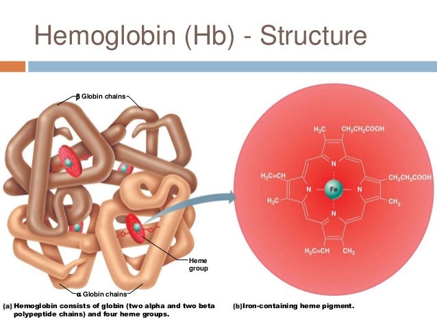
Thalasemia is a genetic condition where the body either does not make enough hemoglobin or the hemoglobin it makes is not normal. Hemoglobin is a component of red blood cells that helps spread oxygen throughout the body. To make hemoglobin the body needs both alpha-globin and beta-globin.
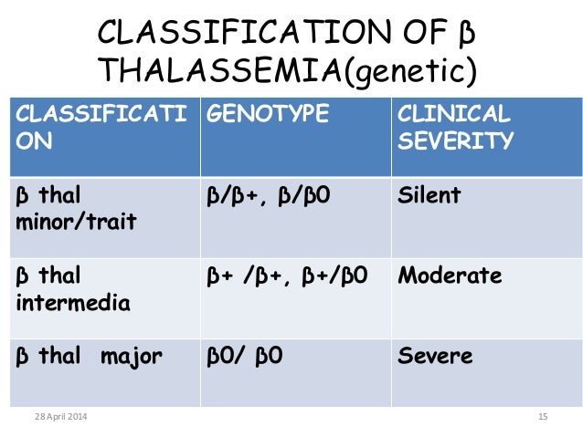
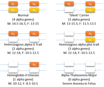
Alpha thalasemia occurs if there are problems with some or all of the alph-globin genes. Beta thalasemia occurs when there are problems with one or both of the beta-globin genes.
Here is a link to an article about thalassemia with lots of info.
http://www.webmd.com/a-to-z-guides/thalassemia-topic-overview

 Read more @ http://www.merckmanuals.com/professional/hematology-and-oncology/anemias-caused-by-hemolysis/thalassemias
There are 2 types of globins in a rbc: alpha & beta globin.
Thalasemia is a genetic condition where the body either does not make enough hemoglobin or the hemoglobin it makes is not normal. Hemoglobin is a component of red blood cells that helps spread oxygen throughout the body. To make hemoglobin the body needs both alpha-globin and beta-globin.
Alpha thalasemia occurs if there are problems with some or all of the alph-globin genes. Beta thalasemia occurs when there are problems with one or both of the beta-globin genes.
Here is a link to an article about thalassemia with lots of info.
http://www.webmd.com/a-to-z-guides/thalassemia-topic-overview

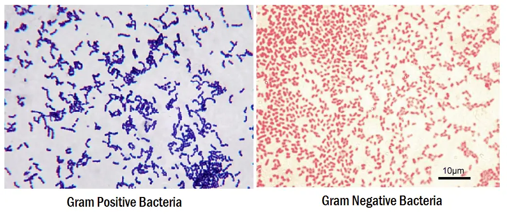
Gram-positive bacteria are bacteria that give a positive result in the Gram stain test, which is traditionally used to quickly classify bacteria into two broad categories according to their cell wall.
Gram-positive bacteria take up the crystal violet stain used in the test, and then appear to be purple-coloured when seen through a microscope. This is because the thick peptidoglycan layer in the bacterial cell wall retains the stain after it is washed away from the rest of the sample, in the decolorization stage of the test.
Gram-negative bacteria cannot retain the violet stain after the decolorization step; alcohol used in this stage degrades the outer membrane of gram-negative cells making the cell wall more porous and incapable of retaining the crystal violet stain. Their peptidoglycan layer is much thinner and sandwiched between an inner cell membrane and a bacterial outer membrane, causing them to take up the counterstain (safranin or fuchsine) and appear red or pink.
Penicillin Bound to Penicillin Binding


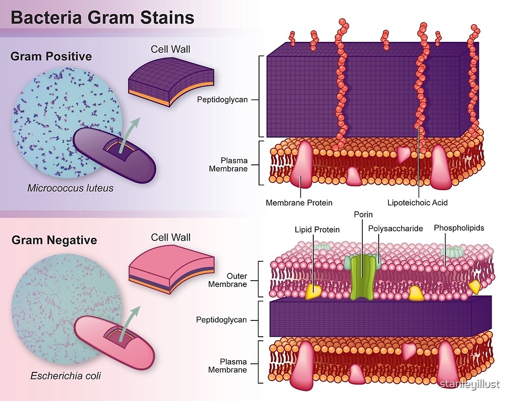


Gram-positive bacteria take up the crystal violet stain used in the test, and then appear to be purple-coloured when seen through a microscope. This is because the thick peptidoglycan layer in the bacterial cell wall retains the stain after it is washed away from the rest of the sample, in the decolorization stage of the test.
Gram-negative bacteria cannot retain the violet stain after the decolorization step; alcohol used in this stage degrades the outer membrane of gram-negative cells making the cell wall more porous and incapable of retaining the crystal violet stain. Their peptidoglycan layer is much thinner and sandwiched between an inner cell membrane and a bacterial outer membrane, causing them to take up the counterstain (safranin or fuchsine) and appear red or pink.
Despite their thicker peptidoglycan layer, gram-positive bacteria are more receptive to antibiotics than gram-negative, due to the absence of the outer membrane (1)
Penicillin is effective only against Gram-positive bacteria because Gram
negative bacteria have a lipopolysaccharide and protein layer that
surrounds the peptidoglygan layer of the cell wall, preventing penicillin from attacking.
Penicillin kills bacteria by inhibiting the proteins which cross-link peptidoglycans in the cell wall
•When a bacterium divides in the presence of penicillin, it cannot fill in
the “holes” left in its cell wall.
•The bacterium is so filled with solutes compared to the surrounding solution
that the water rushes in, and without a full cell wall to support the bacterium,
it “pops” from the turgor pressurePenicillin Bound to Penicillin Binding
Protein 4 (PBP4) Penicillin (shown in the center of the protein as CPK and ball and stick format) acts on a group of proteins (in this case, PBP4) that help form new bacterial wall (3)
References:
1 - https://en.wikipedia.org/wiki/Gram-positive_bacteria
2- Medical microbiology blog: https://blogger.googleusercontent.com/img/b/R29vZ2xl/AVvXsEg_WYvslNVxhMBLGx7WB6d_-9x-sQ2UQDVwVsEz8l-1aI6VRev9TrYk0ksq8CECnkUWWEvcfVnXssI6D5YIEbtN_NthumjfQyBa1TgNqvDrV9rMmh4Fn7EE-Zdg6O3oge0L0JLGQLgOUMU/s1600/Gram-Staining-Procedure.jpg
3 - http://cbm.msoe.edu/includes/pdf/smart2007/mwp2007.pdf