Showing posts with label bf. Show all posts
Wonder Me!
Do NOT confuse Alpha Thalessemia with Beta Thelassemia. Even tho Thalassemia minor is MOST similar to Alpha trait, the number of alleles affected is different in each condition.

If you have too little Alpha to make your rbc, you'll pack it with lots of Beta - which is abnormal.

Hg H disease bc in electrophoresis that's the most H, not A as in normal Hgb electrophoresis.

Usu. looking at lab with normal rbc but microcytic, can kinda make  out that it's Thalessemia, electrophoresis serves little if any purpose other than genetic counseling later on. 

Most commonly seen in adult clinicals: Trait Alpha thalassemia
Hg H & Alpha Thalassemia Major: usu. already dx in peds or neonatology or Maternal Fetal Medicine

Trait: more problems if too heavy menstruation.

Alpha Major: usu. very bad, already

History: yeah I was told I had some type of anemia but I've never had any symptoms.

Lab: usu. normal RBC, low Hgb, Low MCV (microcytic anemia) however rbc is normal bc the body is able to make enuf rbc with the Hgb & iron on hands. Whereas iron deficiency anemia, the body is unable to make enuf rbc due to not even having enuf iron to even make it.


~by Qt
Wonder Me!
Thalessemia is a defective globin (as in heme-o-globin) disorder.
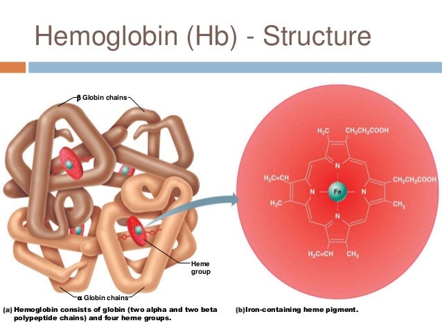
There are 2 types of globins in a rbc: alpha & beta globin.

Thalasemia is a genetic condition where the body either does not make enough hemoglobin or the hemoglobin it makes is not normal. Hemoglobin is a component of red blood cells that helps spread oxygen throughout the body. To make hemoglobin the body needs both alpha-globin and beta-globin.
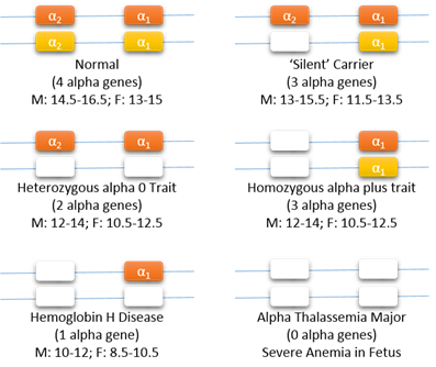
Alpha thalasemia occurs if there are problems with some or all of the alph-globin genes. Beta thalasemia occurs when there are problems with one or both of the beta-globin genes.
Here is a link to an article about thalassemia with lots of info.
http://www.webmd.com/a-to-z-guides/thalassemia-topic-overview


Read more @ http://www.merckmanuals.com/professional/hematology-and-oncology/anemias-caused-by-hemolysis/thalassemias
Wonder Me!
Syncope is transient loss of consciousness due to lack of blood flow to the brain. Usually due to afib with ventricular tachycardia, wolfe parkinson white syndrome.
Labels: 0 comments | | edit post
Wonder Me!
G+: Staphyloccus & Streptococcus (coccus: balls, Strep: chain of balls, Staph: grapes of balls)
G-: Rod
Atypical: Legionella, Chlamydia, Mycoplasma (Macrolides)

Bacteria is usually easy to kill because they can easily be invaded with meds that flood them whereas virus is harder to kill bc of their lack of cells (non-living organisms).


Wonder Me!
B cell is a type of lymphocyte (hence, lymphatic system) which produce antibodies.
Wonder Me!
Protocol: If X, then Y
Learn: If X, why Y? If X, why not Y?
=> Wisdom (lots of learning)

Listen to another presenters => Need to understand what to do next. 
Labels: , 0 comments | | edit post
Wonder Me!
Look for why they were in the ER, (initial cc, final dx)
ER & outpt: cc vs Hospital: why pt is admitted (dx for being admitted) - not cc per se
(due to many different providers: primary care => ER => hospital)

Ex:  Cc:  CP but admitting dx N-STEMI and final dx is still N-STEMI

Ex: Cc:cough & back pain but admission reason: large pleural effusion & hypoxia and find final dx: mets ca. 

=> need summarized view of the assessment of each step 
=> Good example for each step

Also, how long the pts been in the hospital & any hospital procedures
Also, any other details

Ex: Cc cough & back pain x couple months. Admitted for newly found pleural Jeffsion. Final Dx was mets ca. not sure what type. Hospital effar 3 days. Pt got thoracentesis which removed 1.5L of fluid and pt o longer has SOB. Pt got nod on Percocet. And, now they are here.

Ex: Initially presented to ER for CP, admitted for NSTEMI. Only stayed for a day for telemetry observation, she did have a PICC line placed due to poor IV access but no cath lab done. And, we're seeing her today. => No need to mention Echo because it doesn't belong here. It belongs to the Physical Exam section. There will be many tests done for pts with CP - but only mentions ones that will change the course of action. 

Ex: We're seeing her today because she's post d/c. She's 79 y/o Asian Female. Initially, presented to ER for CP, admitted for STEMI.

Ex: Initially presented to ER for CP, admitted for NSTEMI. Only stayed for a day for telemetry observation, she did have a PICC line placed due to poor IV access but no cath lab done. Echo done shows Ej 49%. And, we're seeing her today.=> Placed Echo here because pt's diagnosed with HF.


ROS: Denies CP, weight loss, hematuria, hematemesis, blood in stool or change in bowel habits. => you can say things that you ask. ROS is sth that student is meant to do because it's comprehensive. In real world, we don't have time to be that comprehensive to get that ROS. Often, ROS doesn't give u good info. or it gives u too much info. However, ROS is important in certain situations such as billing. If you bill for a complicated pt, you need to do all ROS. ROS is mostly useful when u're taking care of a pt for whom you don't know anything about. PCP clinic and it's the first time u see the pt, then u may do a more comprehensive ROS. Then, you need to see if tehre's anything there that you need to do to suggest to see if there's anything there taht's not already adddressed or already told. In real practice, not much time to do it except for PCP where you see the pt for the 1st time. In most presentation, you don't tell all ROS. However, you will tell anything that is pertinently pertinent.

Ex: ROS Denies CP, SOB, swelling, edema, or cough but admits difficulty breathing when lying flat  => pertinent data

 The point of presentation is for you to have a structured presentation to collect data. If not, you just do whatever, you'll miss a lot of info. Some will do PE and missing info. This is to remind you of what to do. => Communicating to this other person what you know => structured presentation to make sure comprehensive info and that person has the outline to understand the info. they're getting and what they're going to do next.  => Harder to miss things + How to collect and intepret data + Communicate data

PE: Go from Head to toe => won't forget what to do as the pt's body is our outline => you don't have to remember anything as their body tells you what to do (their physical body) => if I look at this, I'll see this, and this.
General Appearance Pt appears to be in pain curling up in the bed with head up with daughter by bedside.
Mental status  Alert & Oriented to examiner, unable to talk much due to generalized tiredness but able to be very attentive and responds to questions and examination maneuvers (very important unconscious vs can have a conversation, nice person to talk to => massive difference in how we deal with this pt => give general idea of what this pt is like.).
VS

H-E-E-N-T
Lab data
Hypokalemia 1.5 (hospital) => 3.2 (upon nursing home admission)
Hyponatremia 132 (upon nurisng home admission)
INR 1.1 normal
PTT normal
PT high
BS 113
Wbc high
Hct low
Hgb low

Assessment & Plan
 place back on hospice
Morphine for symptom relief


CHF
Wonder Me!

CHF is caused by fluid overload in the heart and. CHF is totally different from COPD. COPD is all about the lungs. Why is it called congestive heart failure? Because it's so much fluid. What kills HF pt? How does it kill the pt? It's the hypoxia. The real immediate problem is hypoxia, not fluid overload. It's not how much fluid int the body but it's how much fluid in the lungs. When the pt is having HF, that's the main problem. Why is there fluid in the lungs? Congestion means it's referring to the lungs. If you do a CXRAY, you'll see big heart. What's congestion? What do you see on cxray? You'll see big heart but that's not important, what's important? FLUID IN THE LUNGS. You'll see pulmonary edema = fluid overload/cushiness/leaking out in the lungs. Pulmonary edema  = that's what you'll see. When CHF pt comes in, the real problem is hypoxia. You can help solve that and makes that better by giving oxygen. But ultimately, you also have to know the root cause.  And, the root cause or the cause is pulmonary edema. PULMONARY EDEMA. So, what we need to know is then to get the fluid out of hte lungs. When the pt is SOB and hypoxic, do you care about getting the fluid out of the legs? NO, you care about getting the fluids out of the lungs: you want to get the pt's legs down ont he ground. Is it going to get their fluid in the legs worse? Yes. What's the problem? The LEFT ventricle. The left ventricle takes blood from the lungs and sends to the rest of the body. The right ventricle tkes blood from the rest of the body and sends it to the lungs. So, CHF, we have blood going to the lungs but we can't pump it out. There's fluid in lungs. Our main problem is the Left ventricle is not strong enough to pump fluid out. So if pt has CHF, FLUID IN THE LUNGS = HYPOXIC. And, if I make the Rt ventricle stronger, will that make the problem better or worse? worse. They may have some weakness in the Rt ventricle but the main problem is the Left ventricle is not effective enuf to pump the blood out. So, the way we can treat that is that we can make the heart stronger. This is a temporary fix. Slowing the gas pedal when the engine is already not doing so well. So, the meds like digoxin and dopamine and dobutamine will stimulate the heart and cause the heart to pump more out. If the heart is okay, it will regulate itself and it will try to push the left side harder to get that fluid out. So, how do we address it otherwise? We give them meds that cause pulling blood in the legs? Stand them up, put the legs down, use gravity. You can give the NTG. What does NTG do? It's a vasodilator. If I vasodilate my peripheral vein, what does that do? I pull more fluid out. It will only go so far tho. Bc you didn't get rid of any fluid in the body but you just move it around to somewhere else. How do I get the fluid out of the body? You actually have to use diuretics. If there's less fluid in the body, there's more fluid going to the Rt ventricle or what? Less. The Rt ventricle is going to pump less. And, the Lt ventricle job easier or harder? Easier.

What's the most common cause of CHF? HTN is a common cause. Sure. What else? too much IV fluid - might be a cause acutely in a pt in a hospital and unable to control their fluid bc their doctor/nurse is giving too much fluid but usuaully not in the world. Why does the pt have a weak heart? Not smoking but smoking makes it worse. Kidney failure? pretty close. CORONARY ARTERY DISEASE due to recurrent MI and hyperlipidemia. The 2 most common causes of CHF = CORONARY ARTERY DISEASE due to recurrent ischemia to the heart and infarction which kill the vessels in the heart and make the heart thin and dilated too large  and weak. The other common cause of CHF in older ppl but not until they're very old is HTN leading to thickening of the heart bc the heart to work hard to push the BP so it gets bigger and thicker and the ventricle cavity smaller. And, they have diastolic or systolic HF? DIASTOLIC. Why diastolic HF? Why? bc it's too thick and it cannot dilate. It's old heart and it's thick. Main issue is poor diastolic function. This is a particular common cause in pt who's older and long-standing HTN. So, if 88 y/o women comes to clinic with HF and she never smokes and she hasn't had many medical problems all her life. She's probably has diastolic HF. If 48 y/o african american man, who eats meat smokes and drinks gambles and does drugs doesn't take care of himself and comes to ur clinic and hospitalized, coronary artery disaese HF, primarily systolic HF bc the heart is too thin so it cannot pump.

PE: edema in legs. Hears fluid in the lungs (crackles).

~bf
Labels: , 0 comments | | edit post