Showing posts with label Cardiology. Show all posts
Wonder Me!


Mechanism

Normally during inspiration, systolic blood pressure decreases by ≤10 mmHg[1] and pulse slightly increases. This is because inspiration makes intra-thoracic pressure more negative relative to atmospheric pressure, which increases systemic venous return to the right atrium by reducing pressure on the veins, particularly the venae cavae. However, the decrease in intra-thoracic pressure and stretching of the lungs during inhalation also expands the compliant pulmonary vasculature so that blood pools in the lungs and decreases pulmonary venous return to the left atrium. Also, the increased systemic venous return to the right side of the heart expands the right heart and directly compromises filling of the left side of the heart by slightly bulging the septum to the left, reducing maximum volume. Reduced left-heart filling leads to a reduced stroke volume which manifests as a decrease in systolic blood pressure, leading to a faster heart rate due to the baroreceptor reflex, which stimulates sympathetic outflow to the heart.
Under normal physiologic conditions the large pressure gradient between the right and left ventricles prevents the septum from bulging dramatically into the left ventricle during inspiration. However such bulging does occur during cardiac tamponade where pressure equalizes between all of the chambers of the heart.[4] Following a zero-sum game principle, as the right ventricle receives more volume it can push the septum into the left ventricle reducing its volume in turn. This additional loss of volume of the left ventricle that only occurs with equalization of the pressures (as in tamponade) allows for the further reduction in volume, so cardiac output is reduced, leading to a further decline in BP. However, in situations where the left ventricular pressure remains higher than the pericardial sac (most frequently from coexisting disease with an elevated left ventricular diastolic pressure), there is no pulsus paradoxus.[5]
Although one or both of these mechanisms may occur, a third may additionally contribute. The large negative intra-thoracic pressure increases the pressure across the wall of the left ventricle (increased transmural pressure, equivalent to [pressure within ventricle] - [pressure outside of ventricle]). This pressure gradient, resisting the contraction of the left ventricle, causes an increase in afterload. This results in a decrease in stroke volume, contributing to the decreased pulse pressure and increased heart rate as described above.
Pulsus paradoxus occurs not only with severe cardiac tamponade, but also with asthma, obstructive sleep apnea and croup. The mechanism, at least with severe tamponade, is likely very similar to those of hypertrophic and restrictive cardiomyopathies (diastolic dysfunction), where a decrease in Left Ventricular (LV) filling corresponds to an increasingly reduced stroke volume. In other words, with these cardiomyopathies, as LV filling decreases, ejection fraction decreases directly, yet non-linearly and with a negative concavity (negative first and second derivatives). Similarly with tamponade, the degree of diastolic dysfunction is inversely proportional to the LV end-diastolic volume. So during inspiration, since LV filling is lesser relative to that during expiration, the diastolic dysfunction is also proportionally greater, so the systolic pressure drops >10 mmHg. This mechanism is also likely with pericarditis, where diastolic function is chastened.

Pulsus Paradoxus = Systolic BP drops during inspiration

Wonder Me!

Cholesterol Charts (what the numbers mean)

Total cholesterol is the sum of all the cholesterol in your blood. Your risk for heart attack and stroke increases with higher cholesterol levels. Other risk factors include smoking, high blood pressure, diabetes, and family history of heart disease or stroke.
Total cholesterol
Less than 200 mg/dL: desirable
200-239 mg/dL: borderline high risk
240 and over: high risk
Wonder Me!
not eating and drinking => Vasovagal syncope
Bridesmaid

Insufficient blood in the brain (fluid insufficiency)
Standing still (blood pooling) 

=> Compression stocking, drinking and eating (don't skip the meal), don't stand still
Wonder Me!
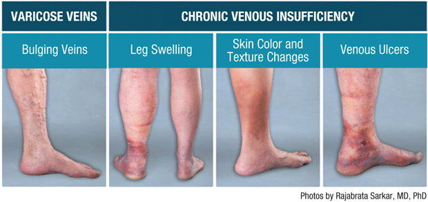
amlodipine (1st line HTN tx) can cause peripheral edema (think bilateral leg swelling)
Wonder Me!

Wonder Me!
Weeping skin erosions

Wonder Me!
Brain natriuretic peptide or B-type natriuretic peptide (BNP) (also ventricular natriuretic peptide or natriuretic peptide B) is a 32-amino acid polypeptide secreted by the ventricles of the heart in response to excessive stretching of heart muscle cells (cardiomyocytes)
ABI
Wonder Me!
ABI Mnemonics:
A => 0.4 (severe) - lower limit
BI =>IB => I.B.=> 1.3 - upper limit
PAD Night => 0.9 (Think you use sanitary pad at night (blood = arterial) for mild-moderate-normal bleeding)
Wonder Me!


Source: Khan Academy, https://youtu.be/_HXl3mu1IoM


 Reversible Causes of Cardiac Arrest: H's and T's

According to The American Heart Association, more than 350,000 people experienced an out of hospital cardiac arrest in 2013. Another 200,000 people had a cardiac arrest in a hospital setting that same year.
Many different traumatic and medical conditions can lead to cardiac arrest in both adults and children. For example, electrical abnormalities, inherited disorders and structural changes in the heart can lead to cardiac arrest. Determining and treating the cause of cardiac arrest is critical to improving patient outcomes. Fortunately, many causes of cardiac arrest are reversible, including the conditions listed below. These conditions are often referred to by the mnemonic “H’s and T’s”:
Hypoxia: Hypoxia is a deficiency in the level of oxygen that reaches the tissues. This problem can occur due to a variety of conditions, such as lung disorders i.e. COPD and asthma. The condition can be reversed by administering oxygen either through BiPAP, a mechanical ventilator, or oxygen mask if the patient has spontaneous respirations.
Hypovolemia: One common cause of cardiac arrest is hypovolemia, which can develop due to excessive fluid or blood loss. It can occur as a result of extreme sweating, severe diarrhea and/or vomiting, or traumatic blood loss. Severe burns can also lead to hypovolemia. After initiating CPR, an intravenous line should be established if possible. The condition is reversed by administering fluids and blood products.
Hypothermia: Although not as common as other causes, hypothermia can also lead to cardiac arrest. When the body’s core temperature drops below 30 degrees Celsius, cardiac output is decreased, which can lead to cardiac arrest. The body may not respond to CPR and defibrillation during the hypothermic state, thus rewarming should be implemented as soon as possible. Depending on how low the body temperature is, passive external rewarming or active internal rewarming may be indicated.
Hypokalemia/Hyperkalemia: Potassium is an electrolyte which plays a role in maintaining normal contraction of the myocardium. If levels become too high or too low, cardiac arrest may ensue. Causes of hypokalemia include excessive vomiting/diarrhea or use of diuretics. Chronic kidney disease can also lead to potassium loss. Treatment may include a controlled but rapid infusion of potassium. Hyperkalemia may be caused by kidney disease, diabetes and as a side effect of certain drugs. Hyperkalemia can be treated by administering sodium bicarbonate or calcium chloride or by performing dialysis.
Hydrogen Ion (Acidosis) Acidosis can be either metabolic or respiratory. Either cause can lead to cardiac arrest. An arterial blood gas is a quick and accurate method to determine if a patient is acidotic. If a patient has respiratory acidosis, he can be treated by providing adequate ventilation. Metabolic acidosis may be treated by administering sodium bicarbonate.
Tension Pneumothorax: A tension pneumothorax develops when there is a buildup of air in the pleural space. The buildup causes a shift in the mediastinum and venous return to the heart is obstructed, which can lead to cardiac arrest. Signs of a tension pneumothorax may include unequal breath sounds, tracheal deviation, difficult ventilation and JVD. The condition can be treated with a needle decompression and/or insertion of a chest tube.
Tamponade (Cardiac): Another reversible cause of cardiac arrest is cardiac tamponade. The condition occurs when fluid or blood fills the pericardium. The fluid puts pressure on the heart and prevents the ventricles from filling properly. It may be caused by trauma to the chest such as a gunshot wound or by inflammation of the pericardium. A pericardiocentesis or a thoracotomy is needed to remove the fluid.
Toxins: One of the most common causes of cardiac arrest is ingestion of a toxins, or an overdose of some type of medication or street drug. One sign of cardiac arrest due to a drug overdose is a prolonged QT interval. In addition to supportive care, a reversing agent may be administered. For example, Narcan may be administered to reverse the effects of narcotic overdose.
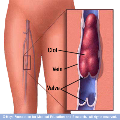
Thrombosis Pulmonary: A pulmonary embolism can lead to cardiac arrest in some instances. A pulmonary embolism usually develops after a blood clot in another area of the body, such as the leg, travels to the pulmonary artery in the lung, leading to cardiac arrest. Prior to the arrest, the patient may exhibit symptoms such as shortness of breath, chest pain, decreased oxygen levels and a cough. Treatment usually includes embolectomy, fibrinolytic therapy or anticoagulant therapy.
Thrombosis Coronary: A coronary thrombosis is a blockage within the coronary artery. It occurs due to blood which has clotted in the vessel. The occlusion in the vessel prevents blood flow to the heart. Cardiac arrest can occur depending on the location and extent of the blockage. Treatment includes angioplasty and stent placement or coronary bypass surgery.
Article Sources
Bakhtiar Ali, MD. Advances in the Acute Management of Cardiac Arrest. The Journal of Emergency Medicine Practice. 2008. Volume 10 (7) https://www.medschool.lsuhsc.edu/emergency_medicine/docs/EMP%20Cardiac%20Arrest.pdf  Accessed August 2014.
Field, JM. Hazinski M, Sayre M. 2010 American Heart Association Guidelines for Cardiopulmonary Resuscitation and Emergency Cardiovascular Care. Circulation. 2010.  http://circ.ahajournals.org/content/122/18_suppl_3/S665.full Accessed August 2014.

Source: https://acls.com/free-resources/pea-asystole/reversible-causes-of-cardiac-arrest-hs-and-ts
Wonder Me!
S/s: Right-sided HF (Tachycardia, decreased O2 saturation, HTN, arrhythmia, hemoptysis, etc.)

Tx:
Vasodilators - Epopros Tenol - Flolan
Soluble guanylate stimulator (Adempas) - 1st and only drug for PAH
CCBs - Sildanefil (Viagra), Tadalafil (Cialis)

Mnemonics: AH-Damn!-GAS (Adempas) & Flow-lane (Flolan)


PAH is a serious disease. This is not to make light of it but picture helps memorization

Ancillary Tx **:
Diuretics - to manage peripheral edema.
Oxygen
Anticoagulants,
Digoxin - improve right ventricular function in patients with right ventricular failure

**Ancillary Tx mnemonics: DO AN


http://www.adempas-us.com/hcp/

https://www.veletri.com/?gclid=CjwKEAiArvTFBRCLq5-7-MSJ0jMSJABHBvp0dai9lBCPRJMB6aNzrC-BTwid9-ZTpdN6ZRj43lPTIhoCNJXw_wcB#isi
Wonder Me!

Symptoms More Likely in Women:

 GI symptoms: indigestion, gas-like pain, dizziness, nausea
Wonder Me!
Generally, HA r/t to HTN is usually generalized due to acute HTN usually r/t marked diastolic BP of rising over 25%

References:
https://books.google.com/books?id=aV1kEf7mlckC&pg=PA435&dq=hypertension+headache+generalized&hl=en&sa=X&ved=0ahUKEwij5pTdnMHSAhVnlFQKHTR9D1gQ6AEIJTAC#v=onepage&q=hypertension%20headache%20generalized&f=false


https://books.google.com/books?id=6n5cAgAAQBAJ&pg=PA59&dq=hypertension+headache+generalized&hl=en&sa=X&ved=0ahUKEwij5pTdnMHSAhVnlFQKHTR9D1gQ6AEIPDAG#v=onepage&q=hypertension%20headache%20generalized&f=false
Wonder Me!
S3, S4:
S3&S4 are lower frequency heart sounds compared with S1&S2. Therefore, it's best heard with the bell of the stethoscope at the apex when pt on the left side in order to bring the heart closer to the surface.

S3
S3 occurs when the chordae tendonae are so tense due to rapid fluid overload filling during diastole. The tense chordae tendonae causes the S3 sound right after the S2 sound (the closure of the semilunar valves).

S3 is normal in children and athletes. It simply means the ventricle is capable of expanding and get more fluid.

S3 is pathologic in middle-aged and older people because it signifies fluid overload (CHF)


S4

S4 occurs when the atrium tries to fill the thick-walled ventricle. This is always pathologic.


References:
S3:
https://books.google.com/books?id=0lxSGJYeXikC&pg=PA36&dq=s3+chordae+tendineae&hl=en&sa=X&ved=0ahUKEwiam5nrt5_SAhVBWmMKHcsHDcIQ6AEIKTAC#v=onepage&q=s3%20chordae%20tendineae&f=false

https://books.google.com/books?id=iAijBQAAQBAJ&pg=PA48&dq=s3+chordae+tendineae&hl=en&sa=X&ved=0ahUKEwiam5nrt5_SAhVBWmMKHcsHDcIQ6AEIMzAE#v=onepage&q=s3%20chordae%20tendineae&f=false
Wonder Me!
Diastolic murmurs: MS/TS AR/PR
Diastolic murmur mnemonics: MS ARD (Mitral Stenosis Aortic Regurgitation Diastole)
Diastolic murmur mnemonics by Qt: MS(TS) AR(PR) D
Diastolic murmurs occur during the diastole phase in which the Atrium is pushing blood from the atrium to the ventricle. This means, during the normal diastole, the M valve will stay open completely while the A valve is closed completely. Hence, murmur occurs when M valve fails to open completely (MS/TS) or the A valve fails to close completely (AR/PR). 


AORTIC REGURGITATION


Aortic regurgitation occurs when the blood flow backwards. Note that in Mitral valve, the normal blood flow is from atrium to ventricle. In mitral valve regurgitation, the backward blood flow thus means the blood from ventricle to atrium. Similarly, in normal aortic valve, the normal blood flow is from ventricle to aorta. Thus, in abnormal aortic valve regurgitation, the blood flows backwards from the aorta to the ventricle. Note that, during diastole, the pressure in the left ventricle is low allowing the filling of the blood from the atrium to the ventricle. Therefore, the previously pumped blood from the previous systole is high leading to high blood pressure in the aorta. As aortic regurgitation occurs, the high pressure blood leakage of blood from aorta to the ventricle causing a high murmur sound heard. Then, as the ventricle is filling up with blood, the sound of the murmur is less and less audible (descendo). Thus, the term early diastolic murmur.

Note that early diastolic murmur (aortic regurgitation) is heard best in the left sternal border instead of the Aortic 2nd right intercostal space because the left seternal border is where the blood is regurgitating back not the Aortic valve location of the 2nd ICS. 

PULMONIC REGURGITATION
Pulmonic regurgitation is similar to aortic regurgitation in mechanism. The only difference is that it's heard best in the upper left side of the sternal border. Pulmonic regurgitation is also a diastolic murmur; more specifically, it's an early diastolic murmur. 


MITRAL STENOSIS


Mitral stenosis occurs during diastole in which it's supposed to be completely open to get blood flow from the atrium to the ventricle. However, in mitral stenosis, the mitral valve is too stenotic to allow blood flow from the atrium to the ventricle. 

It takes the atrium to build up some more pressure than normally to force open the stenotic mitral valve. Thus, it will open loudly (Opening Snap) as the mitral valve is 'cracked' open instead of nicely quietly open in normal valves. The onset of the the opening of the mitral valve is about some msec after the S2 (closure of the A&P valves). 

When the blood coming from the atrium to the ventricle is flowing in, at first, this pressure is very high (recall it takes a lot of pressure to 'crack open' this stenotic valve). Then, when the way is made, the pressure slows down. 

At the last effort, the atrium starts to squeeze all the blood to the ventricle nearing the end of the diastole. This results in the louder sound at the end of the diastole. Thus, it's named 'rumbling sound' at the mid-diastolic. 

Thus, this murmur is termed 'Opening snap + mid-diastolic' with rumbling sound. 

TRICUSPID STENOSIS
Tricuspid stenosis is similar to mitral stenosis: opening snap + mid diaslotic rumbling sound. 


References:
Info & videos mostly from Khan Academy: https://youtu.be/ZUHpAaVpiY8

Wonder Me!
Systolic murmurs:  AS/PS, MR/TR & Mitral Valve prolapse
Systolic murmurs mnemonics: MR. ASS (Mitral Regurgitation Aortic Stenosis Systolic).
Systolic murmurs mnemonics by Qt: MR(TR). AS(PS)S (Remember M&T are 'equivalent' valve and thus will have similar functions/pathology => Remember AP & MT (mnemonics of APT M). Similarly, remember M&T are 'equivalent' valves and thus will have similar functions/pathology => remember from AP&MT (Mnemonics of APT M)

Systolic murmurs:
Normally, during systole (S1 to S2 period), M valve (S1) is supposed to close completely and A valve  (S2) is supposed to open completely. Systolic murmur occurs when M valve fails to close completely leading to an 'eruption 'of blood flowing thru the otherwise closed valve (regurgitation). Systolic murmur also occurs when the A valve fails to open completely leading to leaking of blood flow thru the stenosis valve. Similarly, that's what happens in the T & P valves. ~By Qt.


AORTIC STENOSIS


Aortic stenosis is a systolic murmur (ejection systolic mumur). 

During this time (right after S1), the M valve & the A valve are closed as the pressure is building up in the Lt ventricle. Therefore, there are some msecs until which the the stenotic aortic valve starts to open up. However, since the A valve is stenotic, it won't open all the way creating the murmurs as the blood flows thru the stenotic valve. At first, when the A valve is pushed open, an ejection click (a few miliseconds after the S1) is audible. At this time, blood flow keeps going stronger (crescendo). Then, at a point, the blood flow will be slower gradually (descendo). Thus, the term 'crescendo-descendo' or 'diamond'.

This murmur can also radiate to the neck/carotid as remember that this is an aortic murmur due to aortic stenosis which has blood flows to the carotid

PULMONIC STENOSIS
Pulmonic stenosis is similar to aortic stenosis in terms of mechanism: it's a systolic ejection murmur. The only difference is that it doesn't radiate to the neck (because the blood flow goes to the lungs, evidently). 


 MITRAL VALVE REGURGITATION


Normally, during S1, the M valve will close tightly preventing the backflow (regurgitation) of blood from the Lt ventricle to the Lt atrium. However, due to some pathology, the M valve will not close tightly, thus, termed 'mitral valve regurgitation'. This murmur occurs right when S1 is heard because recall S1 signifies the closure of M valve. M valve is unable to close completely, therefore, the murmur is heard right at S1. 

This regurgitation persists throughout the systole because the blood regurgitates with high pressure from the Lt ventricle to the Lt atrium throughout systole. During diastole (S2 thru S1), the M valve open for blood to flow to the Lt ventricle and you won't hear the regurgitaiton during diastole. Therefore, it's a systolic murmur.

Note that Mitral regurgitation can be heard in the axilla due to anatomic location. 


TRICUSPID REGURGITATION
Tricuspid regurgitation is similar to Mitral regurgitation in terms of mechanism except that you won't hear it in the armpit due to anatomic location.


MITRAL VALVE PROLAPSE 


Mitral valve prolapse occurs when the tendon chordae that anchors the valve to the ventricles become flail and unable to completely close off during systole. Instead, the valve will billow up (prolapse) and cause a mid-systolic click which is non-ejection. It only causes the ejection click in the mid systole because it will take some time to build up enuf pressure to cause a sound heard to ur ears as the pressure builds up in the Left ventricle. 

Sometimes, it can accompany with a mitral valve prolapse when the valve is not only prolapsed but also is unable to open completely. 

Midsystolic click is diagsnotic of Mitral valve prolapse.  


References:
Most information comes from Khan Academy: https://youtu.be/6YY3OOPmUDA 
Some videos & screenshots are from Khan Academy: https://youtu.be/6YY3OOPmUDA


Wonder Me!