Showing posts with label physical signs. Show all posts
Showing posts with label physical signs. Show all posts


Abstract and Introduction
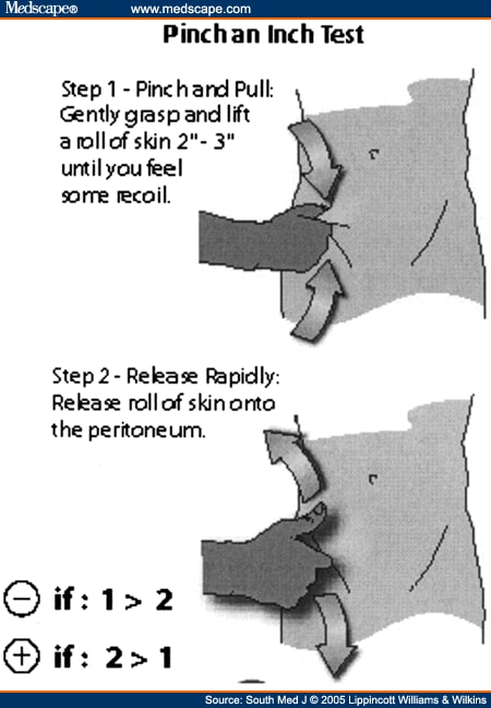
Rebound tenderness is a widely used examination technique for patients with suspected appendicitis, but it can be quite uncomfortable. An alternative test for peritonitis is termed the pinch-an-inch test. This report describes two patients who presented with mild abdominal pain who subsequently were found to have appendicitis. In both patients, classic peritoneal signs were absent, but the pinch-an-inch test was positive. The experienced physician's bedside clinical examination remains the most critical component for rapidly identifying peritonitis. Although rebound tenderness is a widely used examination, it is uncomfortable and may be inaccurate. To perform the pinch-an-inch test, a fold of abdominal skin over McBurney's point is grasped and elevated away from the peritoneum. The skin is allowed to recoil back briskly against the peritoneum. If the patient has increased pain when the skin fold strikes the peritoneum, the test is positive and peritonitis probably is present.
Rebound tenderness, a widely used physical examination test for patients with suspected appendicitis, can be quite uncomfortable for the patient.[1,2] Accordingly, some standard references no longer advise its use on patients with abdominal pain.[3,4] We recently developed an alternative test for peritonitis that in our experience produces less discomfort for patients. We colloquially termed this peritoneal sign the pinch-an-inch test. To the best of our knowledge, others have not described it.
Our pinch-an-inch test is essentially a form of rebound tenderness, only in reverse. To perform the test, a fold of abdominal skin over McBurney's point is grasped and elevated away from the peritoneum (see Fig. 1). The skin is then allowed to recoil back briskly against the peritoneum. If the patient has increased pain when the skin fold strikes the peritoneum, the test is positive and peritonitis is presumably present. As an added feature, if the pain seems excessive just during the initial pinch phase, the patient may have a very low pain threshold, a factor that can be taken into account when deciding if the patient has a surgical abdomen. We anecdotally have found the test to be remarkably helpful for the evaluation of appendicitis as exemplified by the following cases.
Source: http://www.medscape.com/viewarticle/521231
Blumberg sign (or Rebound tenderness positive sign) is elicited by palpating slowly and deeply over a viscus and then suddenly releasing the palpating hand. (2)
Rebound tenderness is tested for by pressing firmly and steadily on a patient's abdomen for a minute or two, and then releasing your hand suddenly. If he finds this agonizingly painful, the sign is positive. It is an uncomfortable and not a very reliable sign, and is most useful when pressure applied in one place causes rebound pain in another. For example, if pressure in his left lower abdomen causes pain in his right lower abdomen, it suggests appendicitis (Rovsing's sign). Many surgeons use light percussion, which is more accurate and much less cruel than rebound tenderness. (1)
1 - http://www.meb.uni-bonn.de/dtc/primsurg/docbook/html/x2980.html
2 - https://offlineclinic.com/blumbergs-sign-rebound-tenderness/
Rebound tenderness is tested for by pressing firmly and steadily on a patient's abdomen for a minute or two, and then releasing your hand suddenly. If he finds this agonizingly painful, the sign is positive. It is an uncomfortable and not a very reliable sign, and is most useful when pressure applied in one place causes rebound pain in another. For example, if pressure in his left lower abdomen causes pain in his right lower abdomen, it suggests appendicitis (Rovsing's sign). Many surgeons use light percussion, which is more accurate and much less cruel than rebound tenderness. (1)
1 - http://www.meb.uni-bonn.de/dtc/primsurg/docbook/html/x2980.html
2 - https://offlineclinic.com/blumbergs-sign-rebound-tenderness/


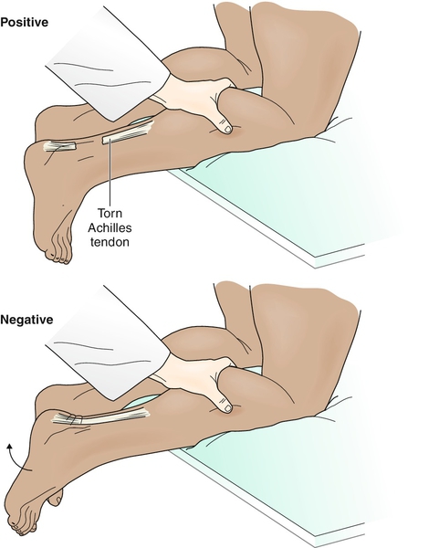
Thompson's test: tests whether the Achille's tendon is intact. If the Achilles' tendon is intact, when the calf muscle is squeezed, the foot will plantar-flex. If the Achilles' tendon is ruptured, the foot will NOT plantar-flexed as the calf muscle is squeezed.
Why it works? The soleus & gastrocnemius make up the calf muscles. Both of these 2 muscles attach to the Achilles' tendon. Therefore, when the you squeeze the calf muscles, you are trying to lift these 2 muscles up contracting the muscles leading to the plantar-flexion of the foot.
Images:


Why it works? The soleus & gastrocnemius make up the calf muscles. Both of these 2 muscles attach to the Achilles' tendon. Therefore, when the you squeeze the calf muscles, you are trying to lift these 2 muscles up contracting the muscles leading to the plantar-flexion of the foot.
Images:
Video:
Just kinda tilt evert and invert to feel how laxity it is for the ankle talus joint




Maisonneuve fracture: Maisonneuve fracture is a fracture in the proximal fibula. It is usually overlooked since patient is more focused on the pain in the ankle than tenderness or pain in the proximal fibula. Check for maisonneuve fx for fx s/s in the ankle. (1)
Why it works: Forceful external rotation & internal rotation of the ankle cause the separation of the distal edge of the fibula. Fibula is a long bone. As the distal edge is pulled away from the tibula, the part near the proximal end endures great stretch. As a lever, the part near the proximal end breaks away from the proximal end causing a fracture. (1)
Case example: http://manuetcorde.org/2013/03/03/dr-maisonneuve-his-fracture-and-why-students-should-learn-their-eponyms/
X-rays: http://radiopaedia.org/articles/maisonneuve-fracture
Mechanism videos:
----
Sources:
(1) https://books.google.com/books?id=YmQ3GGGjDhMC&pg=PA646&dq=maisonneuve+fracture&hl=en&sa=X&ved=0ahUKEwjlzuDXgdDLAhXLJx4KHWGJDM44ChDoAQgpMAM#v=onepage&q=maisonneuve%20fracture&f=false)
Why it works: Forceful external rotation & internal rotation of the ankle cause the separation of the distal edge of the fibula. Fibula is a long bone. As the distal edge is pulled away from the tibula, the part near the proximal end endures great stretch. As a lever, the part near the proximal end breaks away from the proximal end causing a fracture. (1)
Case example: http://manuetcorde.org/2013/03/03/dr-maisonneuve-his-fracture-and-why-students-should-learn-their-eponyms/
X-rays: http://radiopaedia.org/articles/maisonneuve-fracture
Mechanism videos:
----
Sources:
(1) https://books.google.com/books?id=YmQ3GGGjDhMC&pg=PA646&dq=maisonneuve+fracture&hl=en&sa=X&ved=0ahUKEwjlzuDXgdDLAhXLJx4KHWGJDM44ChDoAQgpMAM#v=onepage&q=maisonneuve%20fracture&f=false)
Why it works?: anatomically, squeezing the proximal calf will cause separation of the distal fibula and specifically will cause separation of the anterior tibiofibular ligament
Mechanism of injury: "External rotation and excessive dorsiflexion of the foot on the leg have been reported as the most common mechanisms of injury. The injury is most often incurred by individuals who participate in skiing, football, soccer, and other sport activities played on turf" (2)
"The 3 proposed mechanisms of in-
jury for the syndesmotic injury include: (2)
1 - external
rotation of the foot,9,22,24,26,43
2 - eversion of the talus
within the ankle mortise,22,43 and
3 - excessive
dorsiflexion.9,24,43"
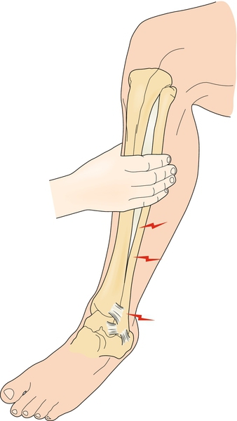
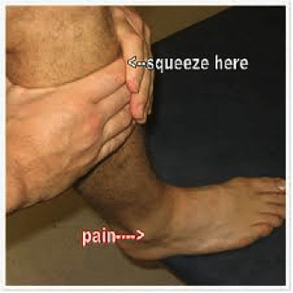


Video: http://www.physio-pedia.com/index.php?title=Squeeze_Test
Syndesmosis joint pain: http://www.physio-pedia.com/Syndesmotic_Ankle_Sprains
(2) http://www.jospt.org/doi/abs/10.2519/jospt.2006.2195
Why it works?: Displaces the anterior talofibular ligament.


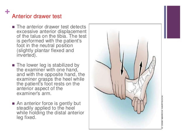


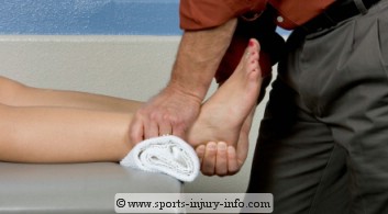

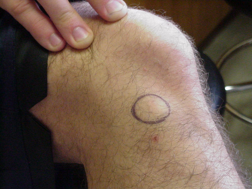


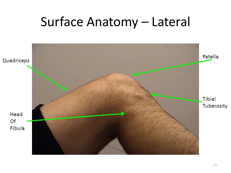


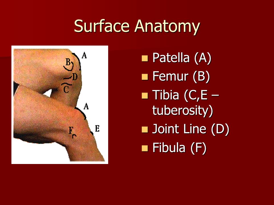


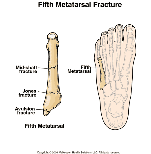
Base of 5th metatarsal bone (lateral view):
Palpation: Palpate the base of the 5th metatarsal bone by palpating along the lateral edge of the foot. About midway between the heel and the fifth little toe lies the tuberosity of the 5th metatarsal bone (3).


Medial malleolus: 6 cm along the edge of the posterior medial malleolus
.jpg)
Navicular (medial surface):
Surface anatomy palpation of the navicular bone: Palpate along the medial side of the foot from the heel, you'll feel navicular prominence anterior to the sustentaculum tali (1), (2).


Sources:
(1) Navicular surface anatomy: http://www.flashcardmachine.com/hoppenfeld-ankle-footpalpation.html
(2) Navicular surface anatomy: https://web.duke.edu/anatomy/mbs/Lab15_MBS/Lab16_preLab.html
(3) 5th metatarsal bone palpation: http://www.dummies.com/how-to/content/surface-anatomy-of-the-knee-and-foot.html
Leg surface anatomy: https://web.duke.edu/anatomy/lab15/lab16_prelab.html
Ankle/foot surface anatomy: https://sites.google.com/site/1styearportfolio/the-ankle-foot/bones-landmarks
Palpation of bony landmarks:
https://www.fgc.edu/wp-content/uploads/2011/12/palpation-of-bony-landmarks.pdf
Ottawa rule for the ankle/foot: http://www.lightspeedphysio.com/ottawa_ankle.png
http://shs-manual.ucsc.edu/sites/shs-manual.ucsc.edu/files/Ottawa%20rules%20for%20x-ray%20of%20ankle%20%26%20foot.pdf
http://www.aafp.org/afp/2001/0101/p93.html
http://www.aafp.org/afp/2002/0901/p785.html
http://www.ncbi.nlm.nih.gov/pmc/articles/PMC1303302/pdf/westjmed00352-0069.pdf
Ottawa rule misses a fracture in the talus from high fall: http://emj.bmj.com/content/18/6/521.full
It is important to understand that Ottawa rules are highly sensitive but not that specific for fractures. Therefore, Ottawa guides you whether to order X-Ray or not. However, Ottawa rules do not tell you whether a fracture is present or not (high sensitivity, low specificity) (2) (5). Therefore, Ottawa rules are helpful to make sure you don't miss a fracture. Yet, Ottawa rules are not made for ruling out fractures. In conclusion, Ottawa rules deem a sensitive test, not a specific test.
Many fractures do not appear on X-Ray. Therefore, absence of fractures on X-Ray does NOT rule out clinical fractures. These fractures include but not limited to (1):
1 - Tibial plateau fx
2 - Segond fx
3 - Salter-Harris type 1
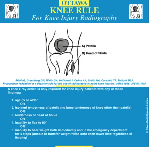
How to use Ottawa rules (3), (4):
(7) Ankle: http://reference.medscape.com/calculator/ottawa-ankle-rule
(8) Foot: http://reference.medscape.com/calculator/ottawa-foot-rule-xray
(9) Knee: http://reference.medscape.com/calculator/ottawa-knee-rule-xray
Isolated tenderness of patella (no bone tenderness of knee other than patella)
---
Sources:
(1) http://www.racgp.org.au/afp/2012/april/the-ottawa-knee-rules/
(2) http://www.jwatch.org/em199910010000001/1999/10/01/ottawa-ankle-rules-miss-some-significant
(3) http://reference.medscape.com/calculator/ottawa-knee-rule-xray
(4) http://www.med.unc.edu/emergmed/education/medical-students/files/Ottawa%20rules%20for%20x-ray%20of%20k...pdf
(5) https://books.google.com/books?id=A2fXLgQ6zQEC&pg=PA99&dq=%22ottawa+rules%22&hl=en&sa=X&ved=0ahUKEwiC5-GVu8vLAhXElx4KHZdADTsQ6AEIIzAB#v=onepage&q=%22ottawa%20rules%22&f=false
(6) Image of Ankle - Ottawa rule: http://www.gpnotebook.co.uk/simplepage.cfm?ID=x20060717214620511440
Many fractures do not appear on X-Ray. Therefore, absence of fractures on X-Ray does NOT rule out clinical fractures. These fractures include but not limited to (1):
1 - Tibial plateau fx
2 - Segond fx
3 - Salter-Harris type 1
How to use Ottawa rules (3), (4):
(7) Ankle: http://reference.medscape.com/calculator/ottawa-ankle-rule
(8) Foot: http://reference.medscape.com/calculator/ottawa-foot-rule-xray
(9) Knee: http://reference.medscape.com/calculator/ottawa-knee-rule-xray
Isolated tenderness of patella (no bone tenderness of knee other than patella)
---
Sources:
(1) http://www.racgp.org.au/afp/2012/april/the-ottawa-knee-rules/
(2) http://www.jwatch.org/em199910010000001/1999/10/01/ottawa-ankle-rules-miss-some-significant
(3) http://reference.medscape.com/calculator/ottawa-knee-rule-xray
(4) http://www.med.unc.edu/emergmed/education/medical-students/files/Ottawa%20rules%20for%20x-ray%20of%20k...pdf
(5) https://books.google.com/books?id=A2fXLgQ6zQEC&pg=PA99&dq=%22ottawa+rules%22&hl=en&sa=X&ved=0ahUKEwiC5-GVu8vLAhXElx4KHZdADTsQ6AEIIzAB#v=onepage&q=%22ottawa%20rules%22&f=false
(6) Image of Ankle - Ottawa rule: http://www.gpnotebook.co.uk/simplepage.cfm?ID=x20060717214620511440
Gray-Turner's sign is a sign that indicates hemorrhage in the peritoneum. It is a bruise seen in the side of the abdomen (flank). When pancreas enzymes got released into the retroperitoneal cavity, it causes bleeding from the retroperitoneum to the subcutaneous tissues of the area between the the end of the rib cage and the ilium (hip bone), aka the flank area (1). Even tho it's commonly thought to be attributed to acute pancreatitis, it can also occurs in AAA (abdominal aortic aneurysm).
Why it works?:
In normal physiology: pancreas releases enzymes that dissolve fat and nutrients in the small intestines. Remember that the pancreas lies behind the stomach and in front of the spine, it is, therefore, a retroperitoneal organ. Also, remember that the pancreas releases enzymes to the duodenum which is the first part of the small intestine that connects directly to the stomach. Therefore, you can image how the pancreas releases enzymes directly into the intestines.
In pancreatitis pathophysiology: pancreatic enzymes destroy the pancreas parenchyma and blood vessels. The damage of the blood vessels causes retroperitoneal hemorrhage.
In pancreatitis, the anatomic pathways of the pancreatic hemorrhage: "hemorrhage originates from the anterior pararenal space => spreading to posterior renal fascia => then to the lateral edge of the quadratus lumborum muscle (flank wall structures).: "The lumbar triangle is a site of anatomic weakness"=> "structural predisposition" to hemorrhaging spreading (according to (2)).
Mnemonics: Grey TURNer’s sign is located when you TURN towards your flank
-----
Sources:
1 - Clinical images of Gray Turner's & Cullen's signs: http://www.nejm.org/doi/full/10.1056/NEJMicm1504339
2- Anatomic pathways: http://www.ncbi.nlm.nih.gov/pubmed/2910743
Peritoneal anatomic pathways: http://pubs.rsna.org/doi/full/10.1148/rg.322115032
Pancreatitis basic info. for patients from Nytimes: http://www.nytimes.com/health/guides/disease/acute-pancreatitis/overview.html
Pancreas basic info. for patients from Hopkins pathology: http://pathology.jhu.edu/pc/BasicOverview1.php?area=ba
Acute pancreatitis basic info.: http://drkupe.blogspot.com/2011/02/acute-pancreatitis.html
Mnemonics: http://www.internalizemedicine.com/2012/01/grey-turners-sign-and-cullens-sign-mnemonic.html
Retroperitoneal organs embryology development: https://academic.amc.edu/martino/grossanatomy/site/Medical/Lab%20Manual/Gastrointestinal/answers/mediastinum11.htm
Retroperitoneal organs anatomy & pathophysiology lecture: http://www.slideshare.net/dhruvtaneja1/reproteritoneum-anatomy-and-pathology
Chronic pancreatitis: http://www.ddc.musc.edu/public/diseases/pancreas-biliary-system/chronic-pancreatitis.html
Why it works?:
In normal physiology: pancreas releases enzymes that dissolve fat and nutrients in the small intestines. Remember that the pancreas lies behind the stomach and in front of the spine, it is, therefore, a retroperitoneal organ. Also, remember that the pancreas releases enzymes to the duodenum which is the first part of the small intestine that connects directly to the stomach. Therefore, you can image how the pancreas releases enzymes directly into the intestines.
In pancreatitis pathophysiology: pancreatic enzymes destroy the pancreas parenchyma and blood vessels. The damage of the blood vessels causes retroperitoneal hemorrhage.
In pancreatitis, the anatomic pathways of the pancreatic hemorrhage: "hemorrhage originates from the anterior pararenal space => spreading to posterior renal fascia => then to the lateral edge of the quadratus lumborum muscle (flank wall structures).: "The lumbar triangle is a site of anatomic weakness"=> "structural predisposition" to hemorrhaging spreading (according to (2)).
Mnemonics: Grey TURNer’s sign is located when you TURN towards your flank
-----
Sources:
1 - Clinical images of Gray Turner's & Cullen's signs: http://www.nejm.org/doi/full/10.1056/NEJMicm1504339
2- Anatomic pathways: http://www.ncbi.nlm.nih.gov/pubmed/2910743
Peritoneal anatomic pathways: http://pubs.rsna.org/doi/full/10.1148/rg.322115032
Pancreatitis basic info. for patients from Nytimes: http://www.nytimes.com/health/guides/disease/acute-pancreatitis/overview.html
Pancreas basic info. for patients from Hopkins pathology: http://pathology.jhu.edu/pc/BasicOverview1.php?area=ba
Acute pancreatitis basic info.: http://drkupe.blogspot.com/2011/02/acute-pancreatitis.html
Mnemonics: http://www.internalizemedicine.com/2012/01/grey-turners-sign-and-cullens-sign-mnemonic.html
Retroperitoneal organs embryology development: https://academic.amc.edu/martino/grossanatomy/site/Medical/Lab%20Manual/Gastrointestinal/answers/mediastinum11.htm
Retroperitoneal organs anatomy & pathophysiology lecture: http://www.slideshare.net/dhruvtaneja1/reproteritoneum-anatomy-and-pathology
Chronic pancreatitis: http://www.ddc.musc.edu/public/diseases/pancreas-biliary-system/chronic-pancreatitis.html
Murphy's sign strongly indicates cholecystitis. However, the absence of Murphy's sign does not rule out cholescytistis (1). You can do this maneuver by having the patient take a deep breath in to push the diaphragm downward as you push your fingers up the lower costal margin in the midclavicular line trying to go below the liver. If patient stops breathing, it is positive, meaning the patient may have cholecystitis (2). Must confirm with lab and imaging.
Why it works: Recall the location of the gallbladder: under the liver @ the midclavicular line. Since inflamed gallbladder (cholescystitis) causes gallbladder tenderness, the gallbladder being sandwiched between the downward pressure produced by the diaphragm and the hand pushing up, this maneuver produces pain and patient will stop breathing in to relieve pressure being pushed down on the poor gallbladder!
See the anatomical position of gallbladder in relation to the costal margin and the liver:
Sources:
1: http://emedicine.medscape.com/article/171886-clinical#b3
2: http://www.turner-white.com/pdf/hp_nov00_murphy.pdf
3: gallbladder image with relation to skeleton, liver & other anatomical regions (highly accurate!): http://www.med.umich.edu/lrc/coursepages/m1/anatomy2010/html/clinicalcases/cholelithiasis/cholelithiasis.html
See the anatomical position of gallbladder in relation to the costal margin and the liver:
Sources:
1: http://emedicine.medscape.com/article/171886-clinical#b3
2: http://www.turner-white.com/pdf/hp_nov00_murphy.pdf
3: gallbladder image with relation to skeleton, liver & other anatomical regions (highly accurate!): http://www.med.umich.edu/lrc/coursepages/m1/anatomy2010/html/clinicalcases/cholelithiasis/cholelithiasis.html
Helpful links:
All body systems:
https://meded.ucsd.edu/clinicalmed/
http://pre.clinicalskills.pitt.edu
Surface anatomy:
https://sites.google.com/site/1styearportfolio/
Newborns:
http://newborns.stanford.edu/PhotoGallery/
Orthopedic/Musculoskeletal physical examination techniques:
http://orthosurg.ucsf.edu/oti/patient-care/divisions/sports-medicine/physical-examination-info/
http://medicine.hsc.wvu.edu/media/16693/resident-ortho-rotation.pdf
http://www.merckmanuals.com/professional/SearchResults?query=Medical+History+and+Physical+Examination+in+Musculoskeletal+Disorders
http://www.orthopaedicsone.com/dashboard.action?atl_token=03bcb29718664ddcfb9fd90f3ef0bb858baf1e21
All body systems:
https://meded.ucsd.edu/clinicalmed/
http://pre.clinicalskills.pitt.edu
Surface anatomy:
https://sites.google.com/site/1styearportfolio/
Newborns:
http://newborns.stanford.edu/PhotoGallery/
Orthopedic/Musculoskeletal physical examination techniques:
http://orthosurg.ucsf.edu/oti/patient-care/divisions/sports-medicine/physical-examination-info/
http://medicine.hsc.wvu.edu/media/16693/resident-ortho-rotation.pdf
http://www.merckmanuals.com/professional/SearchResults?query=Medical+History+and+Physical+Examination+in+Musculoskeletal+Disorders
http://www.orthopaedicsone.com/dashboard.action?atl_token=03bcb29718664ddcfb9fd90f3ef0bb858baf1e21


