Showing posts with label disease. Show all posts
CHF
Wonder Me!

CHF is caused by fluid overload in the heart and. CHF is totally different from COPD. COPD is all about the lungs. Why is it called congestive heart failure? Because it's so much fluid. What kills HF pt? How does it kill the pt? It's the hypoxia. The real immediate problem is hypoxia, not fluid overload. It's not how much fluid int the body but it's how much fluid in the lungs. When the pt is having HF, that's the main problem. Why is there fluid in the lungs? Congestion means it's referring to the lungs. If you do a CXRAY, you'll see big heart. What's congestion? What do you see on cxray? You'll see big heart but that's not important, what's important? FLUID IN THE LUNGS. You'll see pulmonary edema = fluid overload/cushiness/leaking out in the lungs. Pulmonary edema  = that's what you'll see. When CHF pt comes in, the real problem is hypoxia. You can help solve that and makes that better by giving oxygen. But ultimately, you also have to know the root cause.  And, the root cause or the cause is pulmonary edema. PULMONARY EDEMA. So, what we need to know is then to get the fluid out of hte lungs. When the pt is SOB and hypoxic, do you care about getting the fluid out of the legs? NO, you care about getting the fluids out of the lungs: you want to get the pt's legs down ont he ground. Is it going to get their fluid in the legs worse? Yes. What's the problem? The LEFT ventricle. The left ventricle takes blood from the lungs and sends to the rest of the body. The right ventricle tkes blood from the rest of the body and sends it to the lungs. So, CHF, we have blood going to the lungs but we can't pump it out. There's fluid in lungs. Our main problem is the Left ventricle is not strong enough to pump fluid out. So if pt has CHF, FLUID IN THE LUNGS = HYPOXIC. And, if I make the Rt ventricle stronger, will that make the problem better or worse? worse. They may have some weakness in the Rt ventricle but the main problem is the Left ventricle is not effective enuf to pump the blood out. So, the way we can treat that is that we can make the heart stronger. This is a temporary fix. Slowing the gas pedal when the engine is already not doing so well. So, the meds like digoxin and dopamine and dobutamine will stimulate the heart and cause the heart to pump more out. If the heart is okay, it will regulate itself and it will try to push the left side harder to get that fluid out. So, how do we address it otherwise? We give them meds that cause pulling blood in the legs? Stand them up, put the legs down, use gravity. You can give the NTG. What does NTG do? It's a vasodilator. If I vasodilate my peripheral vein, what does that do? I pull more fluid out. It will only go so far tho. Bc you didn't get rid of any fluid in the body but you just move it around to somewhere else. How do I get the fluid out of the body? You actually have to use diuretics. If there's less fluid in the body, there's more fluid going to the Rt ventricle or what? Less. The Rt ventricle is going to pump less. And, the Lt ventricle job easier or harder? Easier.

What's the most common cause of CHF? HTN is a common cause. Sure. What else? too much IV fluid - might be a cause acutely in a pt in a hospital and unable to control their fluid bc their doctor/nurse is giving too much fluid but usuaully not in the world. Why does the pt have a weak heart? Not smoking but smoking makes it worse. Kidney failure? pretty close. CORONARY ARTERY DISEASE due to recurrent MI and hyperlipidemia. The 2 most common causes of CHF = CORONARY ARTERY DISEASE due to recurrent ischemia to the heart and infarction which kill the vessels in the heart and make the heart thin and dilated too large  and weak. The other common cause of CHF in older ppl but not until they're very old is HTN leading to thickening of the heart bc the heart to work hard to push the BP so it gets bigger and thicker and the ventricle cavity smaller. And, they have diastolic or systolic HF? DIASTOLIC. Why diastolic HF? Why? bc it's too thick and it cannot dilate. It's old heart and it's thick. Main issue is poor diastolic function. This is a particular common cause in pt who's older and long-standing HTN. So, if 88 y/o women comes to clinic with HF and she never smokes and she hasn't had many medical problems all her life. She's probably has diastolic HF. If 48 y/o african american man, who eats meat smokes and drinks gambles and does drugs doesn't take care of himself and comes to ur clinic and hospitalized, coronary artery disaese HF, primarily systolic HF bc the heart is too thin so it cannot pump.

PE: edema in legs. Hears fluid in the lungs (crackles).

~bf
Labels: , 0 comments | | edit post
Wonder Me!
it can be normal or it can be severe or bad. A lot of time some atelectasis is normal if you've been in bed all day and not getting around. That's why you need the pt to take a deep breath a couple times before you do the pulse ox. If they've been in bed all day, have them take a couple of breaths to clear out atelectasis. Atelectasis has nothing to do smoking. It's usually due to pt staying in bed all day.
Labels: 0 comments | | edit post
Wonder Me!
COPD usually caused by smoking. COPD = chronic obstructive pulmonary disease. Chronic means >6mo. Obstructive means there's a blockage. Pulmonary means the lungs. So, COPD is the obstructive dz of the lungs. We used to call it emphysema = damage of the alveoli, they are very thin. They don't have much air exchange. They kinda thin out and they get bigger. Lungs is like sponge = tiny little holes and pockets for air to move around. It looks like sponge. It should look like a sponge. COPD lungs are mostly air. And, that's bad. We don't want lungs to be full of air. The lungs should be a sponge. COPD lungs and chest x-ray, it's much more overinflated and clear. U should be able to see it on CXRAY. It's going to look more black.
They also used to be chronic bronchitis. Chronic inflammation of the bronchi. They get narrowed and they get full of junk and they get narrower and they get infxn. They can have some phlegm and coughing and can be like asthma.
When the pt is having a very difficulty time breathing and they try to exhale, it doesn't work. And, that doesn't work. You'll see pt trying to breathe out slowly. They have prolonged expiratory phase. They breathe out very long and slowly with their mouth closed. That's what they call purse lip breathing. They do this to compensate for obstructive bronchi.
Now we know that it's not one thing or another. Most pt have emphysema or chronic bronchitis. Most ppl have features of both. So, hyper-inflated lungs, difficulty breathing, coughing, more infxn, chronic. Do their lungs get oxygen better or worse? Worse? Because damage of alveoli and obstructive bronchi. What's their pulse ox going to be ? Less than normal.

PE: no fluid sounds in lungs (no crackles). but hyper-resonating sounds (egophony).

Labels: 0 comments | | edit post
Wonder Me!
- No dysplasia - 2 EGD w/ biopsy within 1 year
   +If negative, f/u EGD in 3 yrs

- Low-grade dysplasia*
    +Biopsy in 6 months
    +Repeat EGD qyear until no dysplasia x 2

- High-grade dysplasia*
    +Repeat EGD with biopsy q3months + send to ER
    +Surveillance q3months with EGD


*Dx dysplasia should be confirmed with 1 additional pathologist, preferably an expert in esophageal histopathology
Labels: , 0 comments | | edit post
Wonder Me!
Screening for BE may be considered in men with chronic (>5 years) and/or frequent (weekly or more) symptoms of gastroesophageal reflux (heartburn or acid regurgitation) and two or more risk factors for BE or EAC. These risk factors include: age >50 years, Caucasian race, presence of central obesity (waist circumference >102 cm or waist–hip ratio >0.9), current or past history of smoking, and a confirmed family history of BE or EAC (in a first-degree relative) (strong recommendation, moderate level of evidence).


 Given the substantially lower risk of EAC in females with chronic GER symptoms (when compared with males), screening for BE in females is not recommended. However, screening could be considered in individual cases as determined by the presence of multiple risk factors for BE or EAC (age >50 years, Caucasian race, chronic and/or frequent GERD, central obesity: waist circumference >88 cm, waist–hip ratio >0.8, current or past history of smoking, and a confi rmed family history of BE or EAC (in a fi rst-degree relative)). (strong recommendation, low level of evidence).


  Screening of the general population is not recommended (conditional recommendation, low level of evidence).


 Before screening is performed, the overall life expectancy of the patient should be considered, and subsequent

See more:
http://gi.org/wp-content/uploads/2015/11/ACG-2015-Barretts-Esophagus-Guideline.pdf
http://www.medscape.com/viewarticle/854725
Labels: , 0 comments | | edit post
Wonder Me!

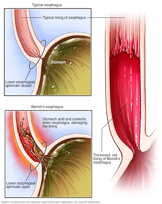
In Barrett's esophagus, normal esophageal cells are replaced with abnormal cells. Barrett's esophagus is thought to be caused by long-standing gastroesophageal reflux disease (GERD), which causes stomach contents to back up into the esophagus.


In Barrett's esophagus, tissue in the tube connecting your mouth and stomach (esophagus) is replaced by tissue similar to the intestinal lining.
Barrett's esophagus is most often diagnosed in people who have long-term gastroesophageal reflux disease (GERD) — a chronic regurgitation of acid from the stomach into the lower esophagus. Only a small percentage of people with GERD will develop Barrett's esophagus.
Barrett's esophagus is associated with an increased risk of developing esophageal cancer. Although the risk is small, it's important to have regular checkups for precancerous cells. If precancerous cells are discovered, they can be treated to prevent esophageal cancer.
Source: http://www.mayoclinic.org/diseases-conditions/barretts-esophagus/basics/definition/con-20027054


Labels: , 0 comments | | edit post
Wonder Me!
Dx test: Anorectal manometry to r/o Hirschsprung's & to aid dx of fecal incontinence.
Labels: , 0 comments | | edit post
Wonder Me!
Source: http://allergycases.blogspot.com/2007/01/mnemonics-asthma.html

Mnemonics: Asthma

Author: V. Dimov, M.D., Allergist/Immunologist and Assistant Professor at University of Chicago
Reviewer: S. Randhawa, M.D., Allergist/Immunologist and Assistant Professor at LSU (Shreveport) Department of Allergy and Immunology

Asthma is the most common chronic respiratory disease, affecting up to 10% of adults and 30% of children (JACI, 2011). Prevalence of asthma is 8%, prevalence of AR is 3 times higher (24%). 40% of patients with AR have asthma, 80% of patients with asthma have AR.

Classification of asthma - mnemonic

I'M MS ("I'm a Master of Science")

Intermittent
Mild persistent
Moderate persistent
Severe persistent

Number of controllers used in each stage of the classification of asthma - mnemonic:

I'M MS
0 1 2 3

0 - SABA PRN (albuterol) only
1 - ICS or LTRA
2 - ICS/LABA or ICS plus LTRA
3 - ICS/LABA and LTRA, consider omalizumab (anti-IgE mAb)

Allergic Rhinitis and its Impact on Asthma (ARIA): Achievements in 10 years and future needs. ARIA has reclassified AR as mild/moderate-severe and intermittent/persistent. This classification closely reflects patients' needs and underlines the close relationship between rhinitis and asthma. http://buff.ly/QL1eYI

Pathogenesis of Asthma

Lymphocytes

CD4, Th2
Central effector cells
Cytokine release

Overview of adhesion molecules, 3 groups remembered by the mnemonic SIS:Selectins
Integrins
Superfamily Ig

Mast cells are subdivided into 2 types based on proteinase content:
TC mast cells -- Tryptase and Chymase in granules
T mast cells -- Tryptase only granules

Mast cells
Mediator release
Mucosal inflammation

Mediators from eosinophils are remembered by the mnemonic CML EEE:

Cytokines
MBP
Lipid Mediators

EDN
ECP
EPO

Eosinophils
Emit
Eight mediators (at least 8, the first C in the mnemonic covers cytokines, chemokines and growth factors)

Overview of adhesion molecules, 3 groups remembered by the mnemonic SIS:Selectins
Integrins
Superfamily Ig

There are 4 families of eicosanoids (PP-LT): prostaglandins (PG), prostacyclins (PGI), leukotrienes (LT) and thromboxanes (TX).

Diagnosis of Asthma

A mnemonic to remember the different PFTs is SPIROMEtry:

Spirometry
PEFR
Inhalation tests:
Reversibilty of
Obstruction with beta-agonist
Metacholine challenge
Exhaled NO

The phases of spirometry can be remembered by the mnemonic BEIF:
Breath normally x 6 times
Exhale fully
Inhalation (deep)
Forceful exhalation for 6 seconds

FEV1/FVC
FEF 25-75
R
Regular (normal) or
Raised in
Restriction

FEV1
1ow in both obstructive and restrictive disease

Bronchodilation test: BB RRBaseline spirometry
Beta-agonist
Repeat spirometry
Reversibilty of obstruction

Methacholine challenge test, remember the numbers: 5-25-20-5:
5 breaths
25 mg/mL metacholine
20% FEV1 reduction
5% of patients with asthma have a negative test, 95% react to the challenge

Test for Respiratory and Asthma Control in Kids (TRACK) 

5
5 questions
5 year-old or younger (2-5 years)

Test for respiratory and asthma control in kids (TRACK) - mnemonic: 3S

Symptoms (3 questions)
SABA use
Steroid use

Test for respiratory and asthma control in kids (TRACK) - complete mnemonic: 3S

Symptoms - SPASymptoms - how often, Play, At night, past 4 weeks
SABA use, past 12 weeks (3 months)
Steroid use, past 12 months (1 year)

Time frame of TRACK:

Symptoms - 4 weeks (1 month)
SABA use - 12 weeks (3 months), quarter
Steroid use - 12 months (1 year)

References:
Test for Respiratory and Asthma Control in Kids (TRACK): A caregiver-completed questionnaire for preschool-aged children. Kevin R. Murphy et al. JACI. Volume 123, Issue 4, Pages 833-839.e9 (April 2009).

Differential Diagnosis of Asthma

C
Children
Congenital conditions
CF

A
Adults
Acquired conditions

Asthma Classification: M MMS

M
ild intermittent

Mild persistent
Moderate persistent
Severe persistent

Treatment

One can remember the stages by the number of controller medications a patient would need at each stage:

I'M MS
0 1 2 3

"Rule of 2s” is used to determine level of control. If any of these are positive, consider a daily controller medication:

- daytime symptoms more than 2 days/wk
- rescue β2 -agonist use more than 2 times per week
- nighttime symptoms more than 2 nights/mo
- more than 2 asthma exacerbations per year
- more than 2 rescue β2-agonist canisters/yr

Reference for rule of 2's: Audio: Asthma, noon conference. Muthiah Pugazhenthi. Podcasting Project for the UT Internal Medicine Residency Program, 12/2006.

If asthma treatment is not working, check DAT:

Diagnosis - not asthma at all (VCD, CF, FBA), asthma plus AR, GERD
Adherence - compliance with medication
Technique - NEB, HFA with spacer, DPI, etc.

3 C's of care - communication, continuity, concordance (finding common ground) are critical for asthma management (http://goo.gl/8gJM6).

Medications

S
Singulair
Single daily dose
Suicude risk (potential)

LABA
M
Monotherapy
Masks inflammation
Mortality increase

Corticosteroids
C category during pregnancy

Budesonide
B category during pregnancy

Exercise-induced asthma treatment: CLIMB

Cromolyn
Leukotriene receptor antagonist
Inhaled steroids
Mast cell stabilizers other than cromolyn
Beta agonists

Leukotriene receptors

Leukotriene
B4
BLT 1, 2 receptors

Leukotriene
C4, D4, E4
CysLT 1, 2 receptors

Ciclesonide mnemonic

C
Ciclesonide 
Converted to active form (des-CIC)
Carboxyl-esterases in bronchial epithelial cells 
Clearance by liver

Published: 01/24/2008
Updated: 11/27/2012
Labels: 0 comments | | edit post
Wonder Me!

Osgood–Schlatter disease (OSD), also known as apophysitis of the tibial tubercle, or Lannelongue's disease,[1] is an inflammation of the patellar ligament at the tibial tuberosity.[2] It is characterized by a painful lump just below the knee and is most often seen in young adolescents. Risk factors include overuse (especially in sports involving running, jumping and quick changes of direction) and adolescent growth spurts.
The condition is named after Robert Bayley Osgood (1873–1956), an American orthopedic surgeon and Carl B. Schlatter, (1864–1934), a Swiss surgeon who described the condition independently in 1903.[1]

Signs and symptoms[edit]

25-year-old male with Osgood–Schlatter disease.
Male with Osgood–Schlatter disease
Osgood–Schlatter disease causes pain in the front lower part of the knee.[3] This is usually at the ligament-bone junction of the patellar ligament and the tibial tuberosity.[4] The tibial tuberosity is a slight elevation of bone on the anterior and proximalportion of the tibia. The patellar tendon attaches the anterior quadriceps muscles to the tibia.[5]
Intense knee pain is usually the presenting symptom that occurs during activities such as running, jumping, squatting, and especially ascending or descending stairs and during kneeling.[citation needed] The pain is worse with acute knee impact. The pain can be reproduced by extending the knee against resistance, stressing the quadriceps, or striking the knee. Pain is initially mild and intermittent. In the acute phase, the pain is severe and continuous in nature. Impact of the affected area can be very painful. Bilateral symptoms are observed in 20–30% of patients.[citation needed]

Diagnosis[edit]

Diagnosis is made based on signs and symptoms.[6]

Ultrasonography[edit]

This test can see various warning signs that predict if OSD might occur. Ultrasonography can detect if there is any swelling within the tissue as well as cartilage swelling.[5] Ultrasonography's main goal is to identify OSD in the early stage rather than later on. It has unique features such as detection of an increase of swelling within the tibia or the cartilage surrounding the area and can also see if there is any new bone starting to build up around the tibial tuberosity.

Types[edit]

Three types of avulsion fractures.
OSD may result in an avulsion fracture, with the tibial tuberosity separating from the tibia (usually remaining connected to a tendon or ligament). This injury is uncommon because there are mechanisms that prevent strong muscles from doing damage. The fracture on the tibial tuberosity can be a complete or incomplete break.
Type I: A small fragment is displaced proximally and does not require surgery.
Type II: The articular surface remains together and the fracture occurs at the junction where the secondary center of ossificationand the proximal tibial epiphysis come together (may or may not require surgery).
Type III: Total break (through articular surface) including high chance of meniscal damage that usually requires surgery.

Differential diagnosis[edit]

Sinding-Larsen and Johansson syndrome,[7] is an analogous condition involving the patellar tendon and the lower margin of the patella bone, instead of the upper margin of the tibia. Sever's disease is a similar condition affecting the heel.

Prevention[edit]

Example of how to stretch the quadriceps muscle.
One of the main ways to prevent OSD is to check the participant's flexibility in their quadriceps and hamstrings. Lack of flexibility in these muscles can be direct risk indicator for OSD. Muscles can shorten, which can cause pain but this is not permanent.[8] Stretches can help reduce shortening of the muscles. The main stretches for prevention of OSD focus on the hamstrings and quadriceps.[9]

Treatment[edit]

Treatment is generally conservative with rest, ice, and specific exercises being recommended.[10] Simple pain killers may be used if required such as acetaminophen (paracetamol) or ibuprofen. Typically symptoms resolve as the growth plate closes.[10] Physiotherapy is generally recommended once the initial symptoms have improved to prevent recurrence.[10] Surgery may rarely be used in those who have stopped growing yet still have symptoms.[10]

Physiotherapy[edit]

Recommended efforts include exercises to improve the strength of the quadricepshamstring and gastrocnemius muscles.[10]
Bracing or use of an orthopedic cast to enforce joint immobilization is rarely required and does not necessarily give quicker resolution. Sometimes, however, bracing may give comfort and help reduce pain as it reduces strain on the tibial tubercle.[11]

Surgery[edit]

Surgical excision may rarely be required in skeletally mature patients.[12] In chronic cases that are refractory to conservative treatment, surgical intervention yields good results, particularly for patients with bony or cartilaginous ossicles. Excision of these ossicles produces resolution of symptoms and return to activity in several weeks. After surgery, it is common for lack of blood flow to below the knees and to the feet. This may cause the loss of circulation to the area, but will be back to normal again shortly. A high pain may come and go every once in a while, due to the lack of blood flow. If this happens, sitting down will help the pain decrease. Removal of all loose intratendinous ossicles associated with prominent tibial tubercles is the procedure of choice, both from the functional and the cosmetic point of view.[13]

Rehabilitation[edit]

Straight leg raises help strengthen the quadriceps without the need to bend the knee. The knee should be kept straight, legs should be lifted and lowered slowly, and reps should be held for three to five seconds.
Rehabilitation focuses on muscle strengthening, gait training, and pain control to restore knee function.[14] Nonsurgical treatments for less severe symptoms include: exercises for strength, stretches to increase range of motion, ice packs, knee tape, knee braces, anti-inflammatory agents, and electrical stimulation to control inflammation and pain. Quadriceps and hamstring exercises prescribed by rehabilitation experts restore flexibility and muscle strength.
Education and knowledge on stretches and exercises is important. Exercises should lack pain and increase gradually with intensity. The patient is given strict guidelines on how to perform exercises at home to avoid more injury.[14] Exercises can include leg raises, squats, and wall stretches to increase quadriceps and hamstring strength. This helps to avoid pain, stress, and tight muscles that lead to further injury that oppose healing. Knee orthotics such as patella straps and knee sleeves help decrease force traction and prevent painful tibia contact by restricting unnecessary movement, providing support, and also adding compression to the area of pain.

Prognosis[edit]

This graph represents the total amount of subjects that were discharged from the hospital with OSD within the years of 2008 and 2011. 100% of patients admitted were discharged, and each year showed that males obtained the disease more frequently than females.
The condition is usually self-limiting and is caused by stress on the patellar tendon that attaches the quadriceps muscle at the front of the thigh to the tibial tuberosity. Following an adolescent growth spurt, repeated stress from contraction of the quadricepsis transmitted through the patellar tendon to the immature tibial tuberosity. This can cause multiple subacute avulsion fracturesalong with inflammation of the tendon, leading to excess bone growth in the tuberosity and producing a visible lump which can be very painful, especially when hit. Activities such as kneeling may also irritate the tendon.
The syndrome may develop without trauma or other apparent cause; however, some studies report up to 50% of patients relate a history of precipitating trauma. Several authors have tried to identify the actual underlying etiology and risk factors that predispose Osgood–Schlatter disease and postulated various theories. However, currently it is widely accepted that Osgood–Schlatter disease is a traction apophysitis of the proximal tibial tubercle at the insertion of the patellar tendon caused by repetitive micro-trauma. In other words, Osgood–Schlatter disease is an overuse injury and closely related to the physical activity of the child. It was shown that children who actively participate in sports are affected more frequently as compared with non-participants. In a retrospective study of adolescents, old athletes actively participating in sports showed a frequency of 21% reporting the syndrome compared with only 4.5% of age-matched nonathletic controls.[15]
The symptoms usually resolve with treatment but may recur for 12–24 months before complete resolution at skeletal maturity, when the tibial epiphysis fuses. In some cases the symptoms do not resolve until the patient is fully grown. In approximately 10% of patients the symptoms continue unabated into adulthood, despite all conservative measures.[12]

Long-term implications[edit]

OSD occurs from the combined effects of tibial tuberosity immaturity and quadriceps tightness.[5] There is a possibility of migration of the ossicle or fragmentation in Osgood-Schlatter patients.[4] The implications of OSD and the ossification of the tubercle can lead to functional limitations and pain for patients into adulthood.[9]
Of people admitted with OSD, about half were children who were between the ages of 1 and 17. In addition, in 2014, a case study of 261 patients was observed over 12 to 24 months. 237 of these patients responded well to sport restriction and non-steroid anti-inflammatory agents, which resulted in recovery to normal athletic activity.[16]

Epidemiology[edit]

Osgood–Schlatter disease generally occurs in boys and girls aged 9–16[17] coinciding with periods of growth spurts. It occurs more frequently in boys than in girls, with reports of a male-to-female ratio ranging from 3:1 to as high as 7:1. It has been suggested that difference is related to a greater participation by boys in sports and risk activities than by girls.[18]

Society and culture[edit]

Steven GerrardRafael NadalPaul ScholesDanny Welbeck and Stephen Ireland are sportsmen who have recovered from this condition.[19] The French tennis player, Gaël Monfils wears patella bands in an attempt to combat the condition.[20]
Labels: 0 comments | | edit post
Wonder Me!
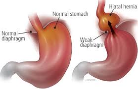
hiatus hernia or hiatal hernia is the protrusion (or herniation) of the upper part of the stomach into the thorax through the esophageal hiatus because of a tear or weakness in the diaphragm. Hiatus hernias often result in heartburn but may also cause chest pain or pain with eating.[1]
The most common cause is obesity. The diagnosis is often by endoscopy or medical imaging.[1]
A hiatus hernia may be treated with lifestyle changes such as raising the head of the bed, weight loss, and adjusting eating habits. Medications such as H2 blockers or proton pump inhibitors may help. If the symptoms do not improve with medications the surgery known as laparoscopic fundoplication may be an option.[1]

Treatment[edit]

In most cases, sufferers experience no discomfort and no treatment is required. If there is pain or discomfort, 3 or 4 sips of room temperature water will usually relieve the pain. However, when the hiatal hernia is large, or is of the paraesophageal type, it is likely to cause esophageal stricture and discomfort. Symptomatic patients should elevate the head of their beds and avoid lying down directly after meals. If the condition has been brought on by stress, stress reduction techniques may be prescribed, or if overweight, weight loss may be indicated. Medications that reduce the lower esophageal sphincter (LES) pressure should be avoided. Antisecretory drugs like proton pump inhibitors and H2 receptor blockers can be used to reduce acid secretion.
Where hernia symptoms are severe and chronic acid reflux is involved, surgery is sometimes recommended, as chronic reflux can severely injure the esophagus and even lead to esophageal cancer.
The surgical procedure used is called Nissen fundoplication. In fundoplication, the gastric fundus (upper part) of the stomach is wrapped, or plicated, around the inferior part of the esophagus, preventing herniation of the stomach through the hiatus in the diaphragm and the reflux of gastric acid. The procedure is now commonly performed laparoscopically. With proper patient selection, laparoscopic fundoplication recent studies have indicated low complication rates, quick recovery, and relatively good long term results.[6][7][8][9][10]
Complications include gas bloat syndromedysphagia (trouble swallowing), dumping syndrome, excessive scarring, and rarely, achalasia. The procedure sometimes fails over time, requiring a second surgery to make repairs.


Labels: 0 comments | | edit post