Showing posts with label ekg. Show all posts
Wonder Me!


Source: Khan Academy, https://youtu.be/_HXl3mu1IoM


 Reversible Causes of Cardiac Arrest: H's and T's

According to The American Heart Association, more than 350,000 people experienced an out of hospital cardiac arrest in 2013. Another 200,000 people had a cardiac arrest in a hospital setting that same year.
Many different traumatic and medical conditions can lead to cardiac arrest in both adults and children. For example, electrical abnormalities, inherited disorders and structural changes in the heart can lead to cardiac arrest. Determining and treating the cause of cardiac arrest is critical to improving patient outcomes. Fortunately, many causes of cardiac arrest are reversible, including the conditions listed below. These conditions are often referred to by the mnemonic “H’s and T’s”:
Hypoxia: Hypoxia is a deficiency in the level of oxygen that reaches the tissues. This problem can occur due to a variety of conditions, such as lung disorders i.e. COPD and asthma. The condition can be reversed by administering oxygen either through BiPAP, a mechanical ventilator, or oxygen mask if the patient has spontaneous respirations.
Hypovolemia: One common cause of cardiac arrest is hypovolemia, which can develop due to excessive fluid or blood loss. It can occur as a result of extreme sweating, severe diarrhea and/or vomiting, or traumatic blood loss. Severe burns can also lead to hypovolemia. After initiating CPR, an intravenous line should be established if possible. The condition is reversed by administering fluids and blood products.
Hypothermia: Although not as common as other causes, hypothermia can also lead to cardiac arrest. When the body’s core temperature drops below 30 degrees Celsius, cardiac output is decreased, which can lead to cardiac arrest. The body may not respond to CPR and defibrillation during the hypothermic state, thus rewarming should be implemented as soon as possible. Depending on how low the body temperature is, passive external rewarming or active internal rewarming may be indicated.
Hypokalemia/Hyperkalemia: Potassium is an electrolyte which plays a role in maintaining normal contraction of the myocardium. If levels become too high or too low, cardiac arrest may ensue. Causes of hypokalemia include excessive vomiting/diarrhea or use of diuretics. Chronic kidney disease can also lead to potassium loss. Treatment may include a controlled but rapid infusion of potassium. Hyperkalemia may be caused by kidney disease, diabetes and as a side effect of certain drugs. Hyperkalemia can be treated by administering sodium bicarbonate or calcium chloride or by performing dialysis.
Hydrogen Ion (Acidosis) Acidosis can be either metabolic or respiratory. Either cause can lead to cardiac arrest. An arterial blood gas is a quick and accurate method to determine if a patient is acidotic. If a patient has respiratory acidosis, he can be treated by providing adequate ventilation. Metabolic acidosis may be treated by administering sodium bicarbonate.
Tension Pneumothorax: A tension pneumothorax develops when there is a buildup of air in the pleural space. The buildup causes a shift in the mediastinum and venous return to the heart is obstructed, which can lead to cardiac arrest. Signs of a tension pneumothorax may include unequal breath sounds, tracheal deviation, difficult ventilation and JVD. The condition can be treated with a needle decompression and/or insertion of a chest tube.
Tamponade (Cardiac): Another reversible cause of cardiac arrest is cardiac tamponade. The condition occurs when fluid or blood fills the pericardium. The fluid puts pressure on the heart and prevents the ventricles from filling properly. It may be caused by trauma to the chest such as a gunshot wound or by inflammation of the pericardium. A pericardiocentesis or a thoracotomy is needed to remove the fluid.
Toxins: One of the most common causes of cardiac arrest is ingestion of a toxins, or an overdose of some type of medication or street drug. One sign of cardiac arrest due to a drug overdose is a prolonged QT interval. In addition to supportive care, a reversing agent may be administered. For example, Narcan may be administered to reverse the effects of narcotic overdose.
Thrombosis Pulmonary: A pulmonary embolism can lead to cardiac arrest in some instances. A pulmonary embolism usually develops after a blood clot in another area of the body, such as the leg, travels to the pulmonary artery in the lung, leading to cardiac arrest. Prior to the arrest, the patient may exhibit symptoms such as shortness of breath, chest pain, decreased oxygen levels and a cough. Treatment usually includes embolectomy, fibrinolytic therapy or anticoagulant therapy.
Thrombosis Coronary: A coronary thrombosis is a blockage within the coronary artery. It occurs due to blood which has clotted in the vessel. The occlusion in the vessel prevents blood flow to the heart. Cardiac arrest can occur depending on the location and extent of the blockage. Treatment includes angioplasty and stent placement or coronary bypass surgery.
Article Sources
Bakhtiar Ali, MD. Advances in the Acute Management of Cardiac Arrest. The Journal of Emergency Medicine Practice. 2008. Volume 10 (7) https://www.medschool.lsuhsc.edu/emergency_medicine/docs/EMP%20Cardiac%20Arrest.pdf  Accessed August 2014.
Field, JM. Hazinski M, Sayre M. 2010 American Heart Association Guidelines for Cardiopulmonary Resuscitation and Emergency Cardiovascular Care. Circulation. 2010.  http://circ.ahajournals.org/content/122/18_suppl_3/S665.full Accessed August 2014.

Source: https://acls.com/free-resources/pea-asystole/reversible-causes-of-cardiac-arrest-hs-and-ts
Wonder Me!


P waves in leads II and V1.
  •      If the P in lead II is greater than 2.5 mV (small boxes) right atrial enlargement (RAE) probably exists. 
  •       If the P wave in V1 is negative or biphasic, then LAE probably exists.  
    • This negative portion of the P wave in V1 should be more than 1 box wide and 1 box down to be considered significant.
PR segment
  • PR depression. The only classic significant PR segment abnormality that is encountered in the emergency department is PR depression.  This is most often seen in the setting of pericarditis.  Since there are multiple stages of pericarditis, these depressions are not always seen when this disease is present.
QRS
  • QRS width:
    • QRS >3ss (0.12 sec) = BBB 


  • QRS shape:
    • RBBB --- RSR' = rabbit ears = The “R” from Right and Rabbit = right BBB (RBBB) there should be an RSR’ in leads V1, V2 or V3.
    • LBBB --- a left BBB (LBBB) you need a deep, wide Q/S wave in these anterior leads.  This you will just have to memorize.


  •  QRS height:
    • LVH --- left ventricular hypertrophy (LVH)
      • A positive deflection in leads I or aVL greater than 11 mV. Mnemonics: 1 and L look like an 11 when they are side by side.
  

      • A value greater than 35 mV when you add the absolute values of the more negative of V1 or V2 plus the more positive of V5 or V6. Mnemonics: just have to look at the Q or S in leads V1 and V2 and see which is more negative.  Take the absolute size of that complex and add it to the larger R of V5 or V6.
      • Remember, only one criterion is sufficient to diagnose LVH.

  • ST segments:
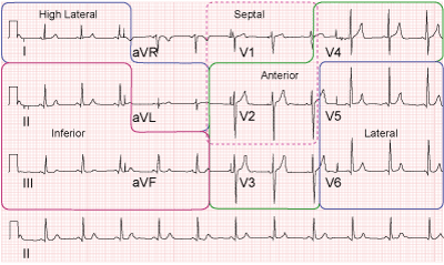
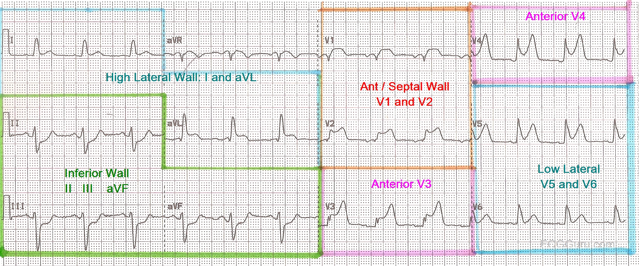
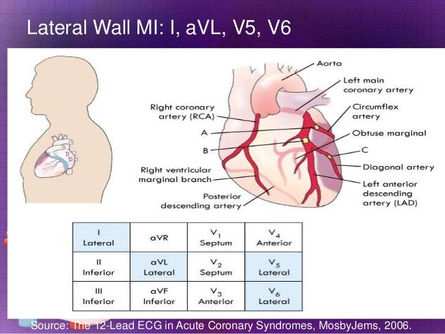
    • you need to check systematically through all 12 leads of the EKG looking for ST elevations or depressions.  These findings are consistent with AMI or ischemia respectively.


  • T wave:
    • Specifically, you are looking for flipped T waves that are pointing in the negative direction.  This is also symbolic of coronary ischemia.  
 
    • Quickly glance at the shape of the T waves.  If they are sharp and pointy instead of nicely rounded, hyperkalemia may exist. 

Labels: 0 comments | | edit post
Wonder Me!
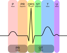
P wave width = 0.12 sec (3ss) (>0.12 or >3ss <=> atrial enlargement)
PR interval (from the beginning of P wave to 1st deflection of QRS) = or <  0.20 sec (5ss = 1ls)  (if >0.20 or 5ss <=> 1st degree AV block)


QRS width = 0.12 sec (3ss) (>0.12 or 3ss <=> bundle branch block (BBB)  (partial or full) OR the complex did not take the conduction pathway at all and had a ventricular origin as is seen in a premature ventricular complex or PVC)

QTc width < 450sec => If >450msec, it may be a tip-off that electrolyte abnormalities exist or some toxin is present.  While this concept is more complex than depicted here, it is not so important and can be discussed later when looking at specific EKG’s


Labels: 0 comments | | edit post
Wonder Me!

Labels: 0 comments | | edit post
Wonder Me!
LAD

Labels: 0 comments | | edit post
Wonder Me!
P wave - precedes every QRS?
P wave morphology - same shape? - originates in every SA node
PR interval - same length?


Labels: 0 comments | | edit post
Wonder Me!

Labels: 0 comments | | edit post
Wonder Me!
Mnemonics 300-150-100-75-60-50
300/n


300/1 = 300
300/2 = 150
300/3 = 100
300/4 = 75
300/5 = 60
300/6 = 50


Labels: 0 comments | | edit post
Wonder Me!
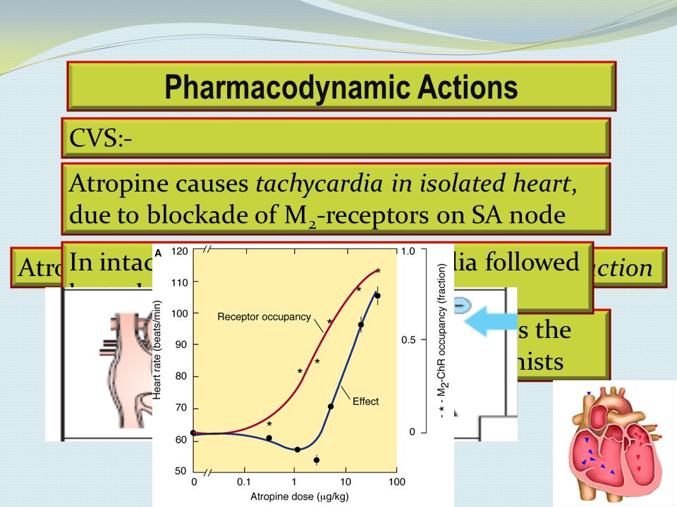
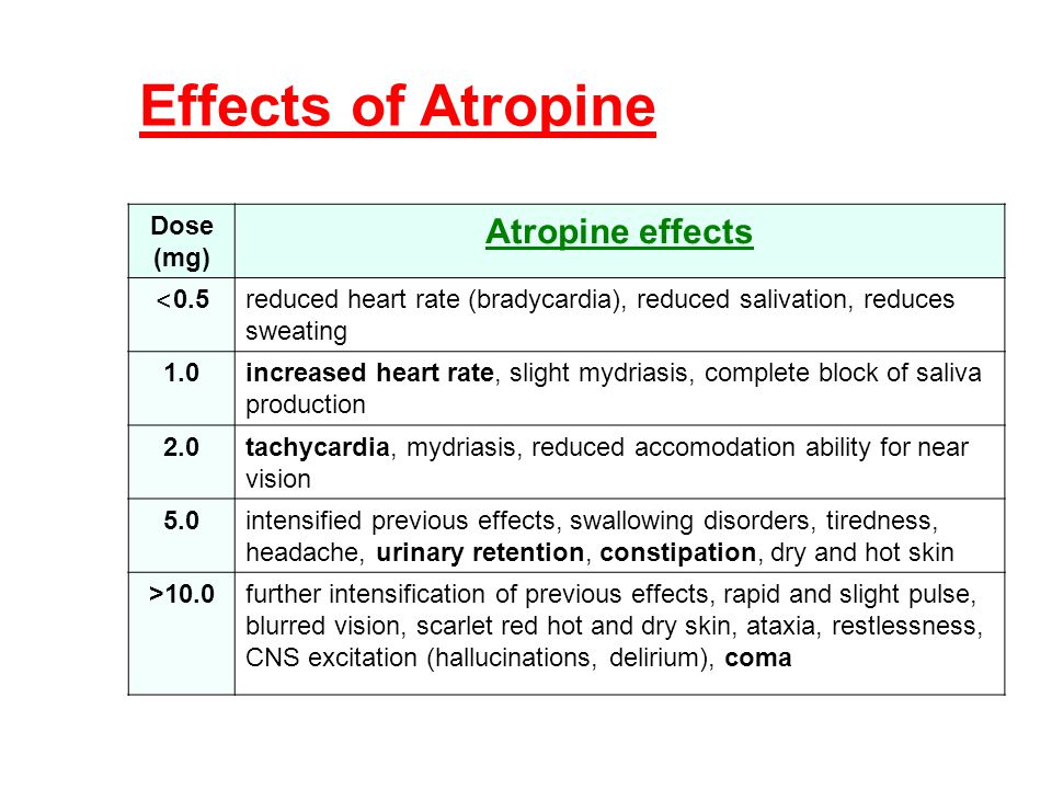
- Atropine is a parasympatholytics that blocks the M2 receptor of the SA node. Therefore, atropine increases the rate of SA node firing & thus => improving the conduction thru the AV node.
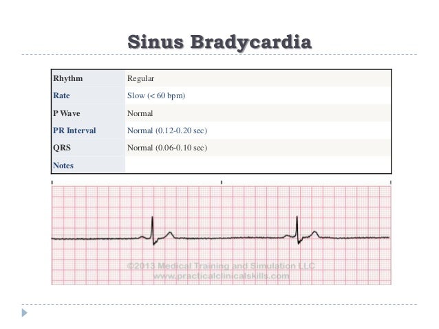
=> Therefore, Atropine can be used for sinus bradycardia because the SA node just needs some kicks.
=> Therefore, Atropine can be used for 2nd degree AV block Wenckebach bc the site of the cause of the AV node wenckeback is at the AV node.
=> Therefore, Atropine is not effective for 2nd degree AV block Mobitz type 2 because the site of the problem blockage is below the AV node.


Labels: 0 comments | | edit post
Wonder Me!


    Causes:
- Electrolyte imbalances
- Acid/Base problem
- Hypoxia
- Temporary or permanent pacing (LOOK FOR THEIR PACEMAKER. If they depend on their pacemaker to fire and it's not working, they probably have permanently slow heart rate! Need the pace maker to be fixed!)

Tx:
None if asymptompatic

If symptomatic (hypotensive, keeps passing out), then:
1 - atropine (atropine is a parasympatholytic, therefore, it will pace the heart making it beat faster; However, be careful not to use atropine on patients with MI because pt will go to V-Tach with the increased myocardial demand workload and oxygen,

2 - pacer, (SA node is not working right, may need pacer)

3 - r/o vagal problem esp. in children bc they like to hold their breath and thus getting into fainting issues a lot!)


Labels: 0 comments | | edit post
Wonder Me!


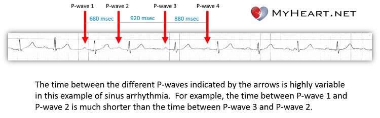
1 - Rate: fast and slow and slow and fast unpredictably

2 - Rhythm:
- P is before each QRS. QRS is after each P.
- Q is normal looking
- Irregular: there's like a pause between some beat

3 - Axis: skip this for now

4 - Width:
- PR interval width is normal (5ss at all beats) and fixed (Remember sinus rhythm has fixed and normal PR interval)
- Q width is normal (<3ss)
- RR: 1/2RR width is less than QT segment= normal

5 - Height:
- No ST elevation
- Upright P wave in lead II (Remember sinus rhythm has upright P wave in lead II)

6 - Pts symptoms:
- Usually EKG sinus arrythmias will have rhythms change as pt breathing in and breathing out
- Usually older pt with syncope, fatigue and worsening HF may have sinus arrhythmias or sinus bradycardia NOT owing to use of beta blocker, that would mean pt may need a pacemaker (sick sinus syndrome)


Labels: 0 comments | | edit post
Wonder Me!
Symptoms: pt not looking at u, totally out, no pulse, no breathing,
EKG:

Tx:
CPR, defib, epinephrine, IV, oxygen, intubation, implantable defibrillator to prevent future incident

Work up: Treatable causes: H's & T's
Labels: 0 comments | | edit post
Wonder Me!
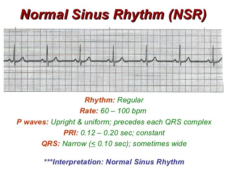
Qt tip:  NSR ALWAYS has upright P wave with normal length PR.
(assuming no dextrocardia or misplaced leads)


Labels: 0 comments | | edit post
Wonder Me!


Labels: 0 comments | | edit post