**All
cases are fictitious. No real patient encounters. Any resemblance to
real persons, living or dead, is purely coincidental.**
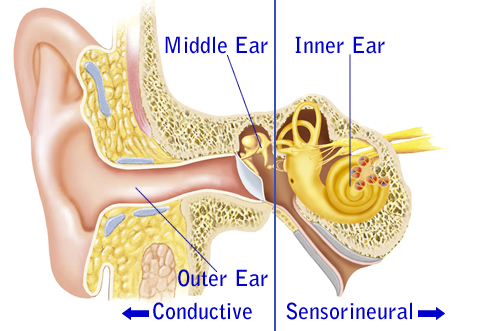
Case*: hmm, I don't see the Tympanic membrane. Did I not push my otoscope far in enough?
Solution:
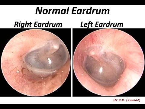
NORMAL TYMPANIC MEMBRANE:
Know what a normal tympanic membrane look like:
- pinkish gray (Wonder me! usually sees normal tympanic membrane more gray thank pinkish)
- small blood vessels along the malleus is normal too
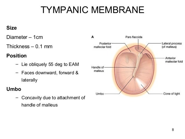
Note: malleus, pars flaccida, pars tensa, & cone of light
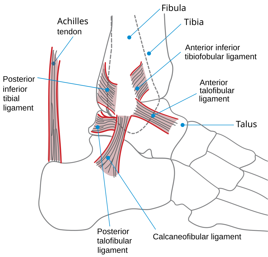
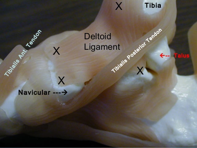
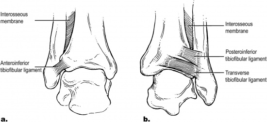
One of the lateral ankle ligaments
Prevents inversion in neutral position
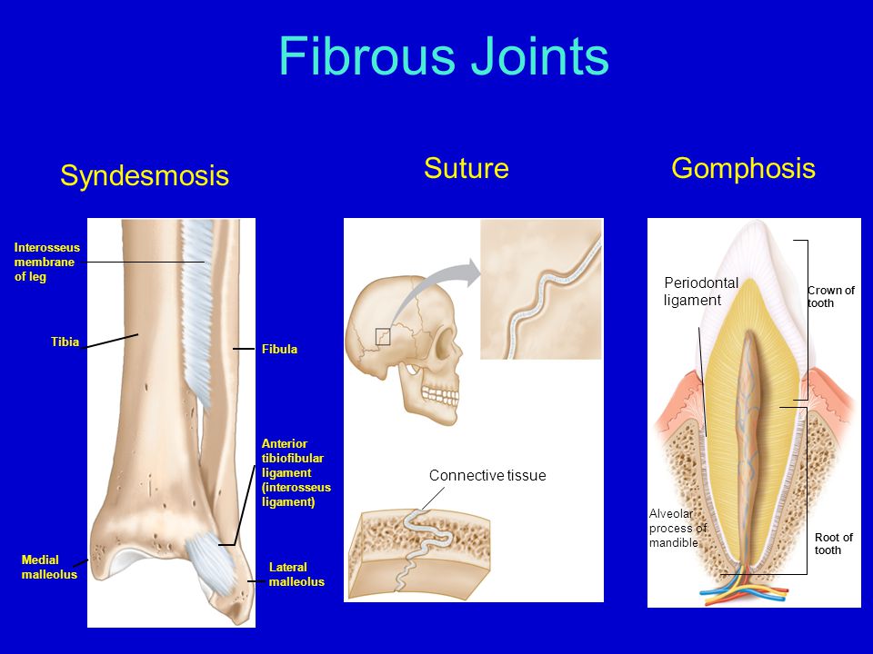
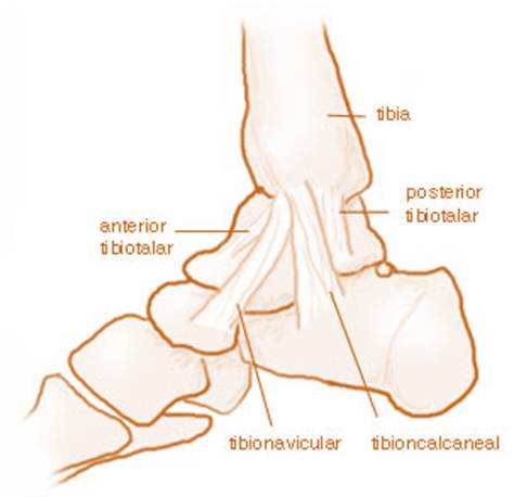
Location: "The calcaneo-fibular ligament is a ligament connecting the apex of the lateral malleolus to the posterolateral aspect of the calcaneus." (1)
Significance: "Tenderness, swelling, and ecchymosis in the area inferior and posterior to the lateral malleolus is evidence of a calcaneo fibular ligament sprain." (1)
One of the ligaments of the lateral ankle - most frequently injured w/ inversion injury.
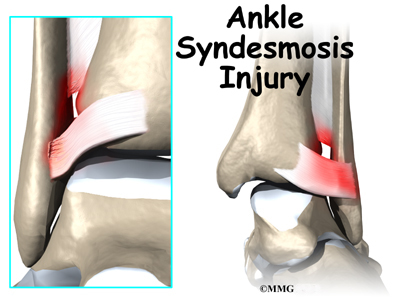
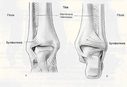
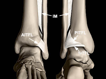
Location: "The talofibular ligament is located between the anterior margin of the lateral malleolus (distal fibula) and the neck of the talus" (1)
Significance: "The talofibular ligament is the most common ankle ligament injured. It is often sprained during an acute ankle inversion. Inspect and palpate the ligament for tenderness, ecchymosis, and swelling to confirm a tear." (1)
This blog claims no credit for any images posted on this site unless otherwise noted. If there is an image appearing on this blog that belongs to you and you do not wish for it appear on this site, please let me know and it will be promptly removed. *All cases are fictitious. No real patient encounters. Any resemblance to real persons, living or dead, is purely coincidental.* Material on this blog is provided for informational purposes only. It is not a substitute for medical care, rehabilitation, educational, consultation, or legal advice. Information on this blog is general as it cannot address each individual’s situation and needs. This blog contains general information which may or may not apply to individuals. This blog cannot and does not address each individual’s situation and needs. I encourage everybody to seek professional advice for any specific questions and concerns. I have made every effort to insure that content is accurate, correct and current and am NOT liable for ANY errors. Images are linked to their websites, please click on the images to be navigated to their original sources. Links to other Websites and contacts have been carefully chosen, but do not imply endorsement and I am NOT responsible or liable for their information and contents. Under NO circumstances, shall the authors and publishers be liable under any theory of recovery for any damages arising out of or in any manner connected with the use of information, services, or documents from the site. Thank you!!!
Quote:
Understanding is key to confidence. Practice helps with psychomotor skills. ~by Qt
Sexy doesn’t impress me. Smart impresses me, strength of character impresses me. But most of all, I am impressed by kindness. Kindness, I think, comes from learning hard lessons well, from falling and picking yourself up. It comes from surviving failure and loss. It implies an understanding of the human condition, forgives its many flaws and quirks. When I see that in someone, it fills me with admiration. ~L.U.












 © My personal NP Prep...
Bloggerized by FalconHive | Wordpress Templates Sponsored by Blogger Styles
© My personal NP Prep...
Bloggerized by FalconHive | Wordpress Templates Sponsored by Blogger Styles