Patients generally have sinusitis or a midface infection (most commonly a furuncle) for 5-10 days. In as many as 25% of cases in which a furuncle is the precipitant, it will have been manipulated in some fashion (eg, squeezing, surgical incision).
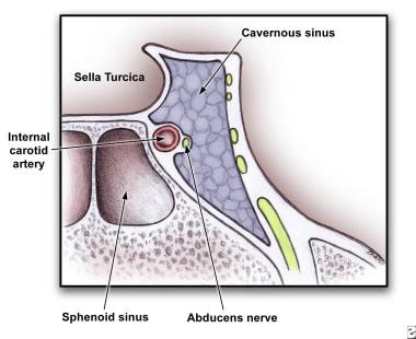
The clinical presentation is usually due to the venous obstruction as well as impairment of the cranial nerves that are near the cavernous sinus.
Headache is the most common presentation symptom and usually precedes fevers, periorbital edema, and cranial nerve signs. The headache is usually sharp, increases progressively, and is usually localized to the regions innervated by the ophthalmic and maxillary branches of the fifth cranial nerve.
In some patients, periorbital findings do not develop early on, and the clinical picture is subtle.
Some cases of CST may present with focal cranial nerve abnormalities possibly presenting similar to an ischemic stroke.[2]
As the infection tracts posteriorly, patients complain of orbital pain and fullness accompanied by periorbital edema and visual disturbances.
Without effective therapy, signs appear in the contralateral eye by spreading through the communicating veins to the contralateral cavernous sinus. Eye swelling begins as a unilateral process and spreads to the other eye within 24-48 hours via the intercavernous sinuses. This is pathognomonic for CST.
The patient rapidly develops mental status changes including confusion, drowsiness, and coma from CNS involvement and/or sepsis. Death follows shortly thereafter.
Tx:
source: http://emedicine.medscape.com/article/791704-clinical#b1
http://patient.info/medicine/metronidazole-for-infection-flagyl
Metronidazole is used to treat a wide variety of infections caused by anaerobic bacteria and micro-organisms called protozoa. These types of organisms often cause infections in areas of the body such as the gums, pelvic cavity and abdomen because they do not need oxygen to grow and multiply. It is commonly prescribed to treat an infection called bacterial vaginosis. It is also prescribed before gynaecological surgery and surgery on the intestines, to prevent infection from developing. Metronidazole can safely be taken by people who are allergic to penicillin.
http://www.stethographics.com/main/physiology_ls_vesicular.html
DYSPHAGIA MNEMONIC (DIFFICULTY SWALLOWING):
Having trouble remembering all the important questions to ask during your patient encounter? Then try this Dysphagia Mnemonic (difficulty swallowing) for USMLE Step 2 CS.
PHYSICAL EXAMINATION for Dysphagia Mnemonic
NOTE: Make sure to wash your hands or wear gloves before you start physical examination. Make sure to ask for permission before you start each physical exam. Make sure to use proper draping(don’t forget to tie back patient’s gown). Make sure to explain each physical examination in layman’s term to your patient. Do NOT repeat painful maneuvers.
- HEENT: Check throat for erythema and exudate
- Neck exam: Check for lymphadenopathy, thyromegaly
- Cardiovascular exam: Auscultation
- Pulmonary exam: Auscultation
- Abdominal exam: Inspection, auscultation, palpation, percussion.
- Skin: check for signs of scleroderma/CREST.
DIFFERENTIAL DIAGNOSIS for Dysphagia Mnemonic
- Esophageal cancer (Dysphagia starts with Solids and progresses to liquids. Hx of chronic alcoholism, smoking & weight loss )
- Achalasia (Dysphasia for BOTH solid and liquids)
- Esophagitis (Pain on swallowing. Immunocompromised “e.g. HIV, Corticosteroids”)
- Systemic Sclerosis (Look for CREST syndrome)
- GERD (Cough at nights, Hoarseness, sore throat)
- Plummer-Vinson syndrome (Iron deficiency anemia, sore throat, craving ice, dirt, clay…)
- Zenker diverticulum (Halitosis, regurgitation)
- Pill-induced esophagitis (e.g. Bisphosphonates)
- Mitral Stenosis (look for an immigrant or a pregnant female)
DIAGNOSTIC WORKUP for Dysphagia Mnemonic
- CBC
- Serum iron, ferritin, TIBC
- Throat culture
- HIV antibody and viral load, CD4 count
- Chest X-ray
- Barium swallow
- Endoscopy
- Esophageal manometry
- Chest CT
http://www.medical-institution.com/dysphagia-mnemonic-difficulty-swallowing-usmle-step-2-cs-mnemonics/
- Bowel Segments
- "Dow Jones Industrial Averages Closing Stock Report" is a good one, even though it misses the Cecum...
Dow
Jones
Industrial
Averages
Closing
Stock
ReportDuodenum
Jejunum
Ileum
Appendix
Colon
Sigmoid
Rectum - Liver Lobes
- The four lobes of the liver: caudate, quadrate, left and right, bring to mind the newspaper headline of the wheelchair bound fellow who left a party right after his ugly girlfriend departed: "QUAD LEFT RIGHT after COW-DATE"
- Pertoneum Facts
- The idea is to relate key letters of related parts...
- stOMach and OMentum (which lays over the stomach)
The bacterium e. coLI is found in the Large Intestine
The OMentum covers the stOMach
The Lesser OMentum holds the Liver and stOMach
The Mesentery holds the sMall intestine
The mesoCOLON attaches the large intestine (COLON) to the posterior abdominal wall.
The periTONEa, which prevents the intestines from kinking, TONES the GI tract. - Sphincters of the Ailmentary Canal
- APE OIL initials the five of them...
A
P
E
O
I
LAnal
Pyloric
(Lower) Esophageal
Oddi
Ileocecum
iLeocecum - Stomach Parts
- "The CAR is FUN 'til the BODY PILES" relates the four parts of the stomach: Cardiac, Fundus, Body, Pylorus. The pylorus is where the food piles waiting for the sphincter to open.
Ulcerative colitis: definition of a severe attack A STATE:
Anemia less than 10g/dl
Stool frequency greater than 6 stools/day with blood
Temperature greater than 37.5
Albumin less than 30g/L
Tachycardia greater than 90bpm
ESR greater than 30mm/hr
Anemia less than 10g/dl
Stool frequency greater than 6 stools/day with blood
Temperature greater than 37.5
Albumin less than 30g/L
Tachycardia greater than 90bpm
ESR greater than 30mm/hr
Vomiting: extra GI differential VOMITING:
Vestibular disturbance/ Vagal (reflex pain)
Opiates
Migrane/ Metabolic (DKA, gastroparesis, hypercalcemia)
Infections
Toxicity (cytotoxic, digitalis toxicity)
Increased ICP, Ingested alcohol
Neurogenic, psychogenic
Gestation
Pancreatitis (acute): causes GET SMASHED:
Gallstones
Ethanol
Trauma
Steroids
Mumps
Autoimmune (PAN)
Scorpion stings
Hyperlipidemia/ Hypercalcemia
ERCP
Drugs (including azathioprine and diuretics)
· Note: 'Get Smashed' is slang in some countries for drinking, and ethanol is an important pancreatitis cause.
IBD: surgery indications "I CHOP":
Infection
Carcinoma
Haemorrhage
Obstruction
Perforation
· "Chop" convenient since surgery chops them open.
Hereditary Nonpolyposis Colorectal Cancer (HNPCC) cause is DNA mismatch repair DNA mismatch causes a bubble in the strand where the two nucleotides don't match.
This looks like the ensuing polyps that arise in the colon.
IBD: extraintestinal manifestations A PIE SAC:
Aphthous ulcers
Pyoderma gangrenosum
Iritis
Erythema nodosum
Sclerosing cholangitis
Arthritis
Clubbing of fingertips
Digestive disorders: pH level With vomiting both the pH and food come up.
With diarrhea both the pH and food go down.
H. Pylori treatment regimen (rough guidelines) "Please Make Tummy Better":
Proton pump inhibitor
Metronidazole
Tetracycline
Bismuth
· Alternatively: TOMB:
Tetracycline
Omeprazole
Metronidazole
Bismuth
Bilirubin: common causes for increased levels "HOT Liver":
Hemolysis
Obstruction
Tumor
Liver disease
Hemolysis
Obstruction
Tumor
Liver disease
Ulcerative colitis: complications "PAST Colitis":
Pyoderma gangrenosum
Ankylosing spondylitis
Sclerosing pericholangitis
Toxic megacolon
Colon carcinoma
Cholangitis features CHOLANGITITS:
Charcot's triad/ Conjugated bilirubin increase
Hepatic abscesses/ Hepatic (intra/extra) bile ducts/ HLA B8, DR3
Obstruction
Leukocytosis
Alkaline phosphatase increase
Neoplasms
Gallstones
Inflammatory bowel disease (ulcerative colitis)
Transaminase increase
Infection
Sclerosing
Charcot's triad/ Conjugated bilirubin increase
Hepatic abscesses/ Hepatic (intra/extra) bile ducts/ HLA B8, DR3
Obstruction
Leukocytosis
Alkaline phosphatase increase
Neoplasms
Gallstones
Inflammatory bowel disease (ulcerative colitis)
Transaminase increase
Infection
Sclerosing
Charcot's triad (gallstones) "Charge a FEE":
Charcot's triad is:
Fever
Epigastric & RUQ pain
Emesis & nausea
Haemachromatosis complications "HaemoChromatosis Can Cause Deposits Anywhere":
Hypogonadism
Cancer (hepatocellular)
Cirrhosis
Cardiomyopathy
Diabetes mellitus
Arthropathy
Pancreatitis: criteria PANCREAS:
PaO2 below 8
Age >55
Neutrophils: WCC >15
Calcium below 2
Renal: Urea >16
Enzymes: LDH >600; AST >200
Albumin below 32
Sugar: Glucose >10 (unless diabetic patient)
Pancreatitis: Ranson criteria for pancreatitis: at admission "GA LAW" (GA is abbreviation for the U.S. state of Georgia):
Glucose >200
AST >250
LDH >350
Age >55 y.o.
WBC >16000
Pancreatitis: Ranson criteria for pancreatitis: initial 48 hours "C & HOBBS" (Calvin and Hobbes):
Calcium < 8
Hct drop > 10%
Oxygen < 60 mm
BUN > 5
Base deficit > 4
Sequestration of fluid > 6L
Pancreatitis: Ranson criteria for pancreatitis at admission LEGAL:
Leukocytes > 16.000
Enzyme AST > 250
Glucose > 200
Age > 55
LDH > 350
GIT symptoms BAD ANAL S#!T:
Bleeding
Abdominal pain
Dysphagia
Abdominal bloating
Nausea & vomiting
Anorexia/ Appetite changes
Lethargy
S#!ts (diarrhea)
Heartburn
Increased bilirubin (jaundice)
Temperature (fever)
Crohn's disease: morphology, symptoms CHRISTMAS:
Cobblestones
High temperature
Reduced lumen
Intestinal fistulae
Skip lesions
Transmural (all layers, may ulcerate)
Malabsorption
Abdominal pain
Submucosal fibrosis
Dysphagia: differential DISPHAGIA:
Disease of mouth and tonsils/ Diffuse oesophageal spasm/ Diabetes mellitus
Intrinsic lesion
Scleroderma
Pharyngeal disorders/ Palsy-bulbar-MND
Achalasia
Heart: eft atrium enlargement
Goitre/ myesthenia Gravis/ mediastinal Glands
Infections
American trypanosomiasis (chagas disease)
21
Dry mouth: differential "DRI":
·2 of each:
Drugs/ Dehydration
Renal failure/ Radiotherapy
Immunological (Sjogren's)/ Intense emotions
Liver failure: decompensating chronic liver failure differential HEPATICUS:
Haemorrhage
Electrolyte disturbance
Protein load/ Paracetamol
Alcohol binge
Trauma
Infection
Constipation
Uraemia
Sedatives/ Shunt/ Surgery
Cirrhosis: causes of hepatic cirrhosis HEPATIC:
Hemochromatosis (primary)
Enzyme deficiency (alpha-1-anti-trypsin)
Post hepatic (infection + drug induced)
Alcoholic
Tyrosinosis
Indian childhood (galactosemia)
Cardiac/ Cholestatic (biliary)/ Cancer/ Copper (Wilson's)
Hemochromatosis (primary)
Enzyme deficiency (alpha-1-anti-trypsin)
Post hepatic (infection + drug induced)
Alcoholic
Tyrosinosis
Indian childhood (galactosemia)
Cardiac/ Cholestatic (biliary)/ Cancer/ Copper (Wilson's)
Hepatic encephalopathy: precipitating factors HEPATICS:
Hemorrhage in GIT/ Hyperkalemia
Excess protein in diet
Paracentesis
Acidosis/ Anemia
Trauma
Infection
Colon surgery
Sedatives
Diabetic ketoacidosis: precipitating factors · 5 I's:
Infection
Ischaemia (cardiac, mesenteric)
Infarction
Ignorance (poor control)
Intoxication (alcohol)
Whipple's disease: clinical manifestations SHELDA:
Serositis
Hyperpigmentation of skin
Eating less (weight loss)
Lymphadenopathy
Diarrhea
Arthritis
Celiac sprue gluten sensitive enteropathy: gluten-containing grains BROW:
Barley
Rye
Oats
Wheat
· Flattened intestinal villi of celiac sprue are smooth, like an eyebrow.
Liver failure (chronic): signs found on the arms CLAPS:
Clubbing
Leukonychia
Asterixis
Palmar erythema
Scratch marks
Clubbing
Leukonychia
Asterixis
Palmar erythema
Scratch marks
Splenomegaly: causes CHIMP:
Cysts
Haematological ( eg CML, myelofibrosis)
Infective (eg viral (IM), bacterial)
Metabolic/ Misc (eg amyloid, Gauchers)
Portal hypertension
Source: http://www.valuemd.com/gastro.php
Source: http://webcache.googleusercontent.com/search?q=cache:7OvEopugpDkJ:unmhospitalist.pbworks.com/f/31.1-24%2520to%25201-30%2520Resident%2520Pancreatitis%2520Module.doc+&cd=20&hl=en&ct=clnk&gl=us
Resident Version
Acute
and Chronic Pancreatitis Module
Created
by Dr. Teodora Konstantinova
Objectives:
1. List two differences in the diagnosis of acute
and chronic pancreatitis.
2. Name 4 risk factors for developing
pancreatitis
3. List two differences between treatment approaches
to acute and chronic pancreatitis
4. Use Ranson criteria to predict severity of
acute pancreatitis.
References:
1. Whitcomb D. C., Acute pancreatitis, N Engl J
Med 2006, 354:2142-2150
2. Steinberg W, Tenner S, Medical Progress:
Acute pancreatitis, NEnglJMed 1994, 330:1198-1210
3. Michael L. Steer, MD, Irving Waxman, MD and
Steve Freedman, MD, Medical progress:
Chronic Pancreatitis, N Engl J Med 1995, 332:1482-1490
4. Ranson JH, Diagnostic Standards for Acute
Pancreatitis World J Surg 1997, 21:136-42
CASE:
HPI: A 60 yo
female presents to the ER with severe mid-epigastric pain with radiation to the
back, nausea and vomiting that started after lunch the previous day. Vomiting did not relieve the pain. The patient also reports coughing with yellow
sputum for about a day. She doesn’t
report fever, but states she has “chills” post emesis.
PMH:
HTN, depression, GERD, and menopausal symptoms requiring hormone
replacement therapy (HRT). The patient
also recently completed a course of nitrofurantoin for cystitis.
PSH: cholecystectomy
6 months ago.
Social hx: Patient
has no history of alcohol abuse.
Medications:
Lisinopril, Paxil, cimetidine, estrogen and completed course of nitrofurantoin
2 days ago.
Physical exam: VS: 100/60, P-
110, R-16, T-37.0 C, P02 - 87 % on RA.
The exam is significant for left lower lung field rales and
egophany. The patient also had moderate
mid-epigastric and RUQ tenderness without rebound or guarding. The rest of the
exam is unremarkable.
Labs:
wbc: 16.5, H/H: 12/45
electrolytes are WNL
SGOT/SGPT: 500/400 U/l, alkaline phosphatase 400 U/l , total Bi 2.0 mg/dL
LDH: 860 IU per
liter, serum lipase-5000, serum amylase- 2000, TG- 300
UA which is neg for LCE, nitrites, bacteria, WBCs or RBCs.
1. At this point in your evaluation, what should
you initially be concerned about?
2. What could be the etiologies of this patient’s pancreatitis?
3. What radiologic studies should you consider
obtaining?
4. Are you able to calculate your patient’s
Ranson’s score?
Discussion Outline:
I. Differences between acute and chronic pancreatitis:
Acute and chronic pancreatitis are distinguished from each
other on the basis of structural and functional criteria.
1. Acute pancreatitis
is an inflammatory condition of the pancreas. Acute disease is characterized by
a normal pancreas that becomes inflamed prior to the attack and once the attack
resolves the pancreas returns to normal.
2. Chronic
pancreatitis is a progressive disorder of the pancreas that causes destruction
of the pancreas. It involves long-term inflammation and scarring of the
pancreas that is irreversible.
II. Pathogenesis:
Acute pancreatitis:
1. It relates to
inappropriate activation of trypsinogen to trypsin (the key enzyme in the
activation of pancreatic zymogens and a lack of prompt elimination of active
trypsin inside the pancreas.
2. Activation of
digestive enzymes causes pancreatic injury and results in an inflammatory
response that is out of proportion to the response of other organs to a similar
insult.
3. The acute
inflammatory response itself causes substantial tissue damage and may progress
beyond the pancreas to a systemic inflammatory response syndrome, multiorgan
failure and death.
Chronic pancreatitis:
1. The hypersecretion
of protein from acinar cells in the absence of increased fluid or bicarbonate
secretion from duct cells is characteristic of chronic pancreatitis.
2. Plugs formed by
the precipitation of protein within the interlobular and intralobular ducts are
an early finding.
3. The plugs contain
multiple proteins (digestive enzymes, glycoproteins, and acidic
mucopolysaccharides).
4. The precipitation
of calcium carbonate in the plugs results in the formation of intraductal
stones (this is more common in patients with alcohol-induced or tropical
pancreatitis).
5. Patients with idiopathic chronic pancreatitis frequently
have an elevated pressure in the pancreatic duct.
III. Causes of pancreatitis:
1. Gallstones (45 %
of cases)
2. Alcohol (35 % of
cases)
3. Drugs:
Azathioprine, Mercaptopurine, Valproic acid, Estrogen, Tetracyclines,
Metronidazole, Nitrofurantion, Pentamidine, Lasix, Sulfonamides, Methyldopa,
Cimetidine, Ranitidine, Salicylates, Erythromycin
4. Metabolic
abnormalities: Hypercalcemia, Hypertriglyceridemia
5. Trauma: accidental
or iatrogenic (ERCP, postoperative, endoscopic sphincterotomy)
6. Infections:
Parasitic (ascariasis, clonorchiasis)
Viral (mumps, rubella, Hepatitis A, B, non-A, non-B,
coxsackievirus B, Echo virus, adenovirus, varicella, Epstein-Barr virus, HIV
virus)
Bacterial: (mycoplasma, Campylobacter
jejuni, Mycobacterium tuberculosis, Legionella, Leptospirosis)
7. Miscellaneous causes: penetrating PUD, Crohn’s disease,
Reye’s syndrome, cystic fibrosis)
8. Idiopathic (10% of
cases)
IV. Diagnosis
Diagnosis of acute pancreatitis:
1. Characteristic
abdominal pain (begins in the upper abdomen and spreads through the back)
2. Nausea and
vomiting (usually the vomiting doesn’t relief the pain)
3. Elevated serum
levels of pancreatic enzymes (amylase and lipase)
4. Both enzymes
remain elevated with ongoing pancreatic inflammation, with amylase level
typically returning to normal shortly before lipase levels in the resolution
phase.
5. Imaging:
-
Ultrasound of the abdomen remains the most sensitive method of
evaluating the biliary tract in acute pancreatitis. It has a sensitivity of 67%
in the urgent diagnosis, and a specificity of 100%.
-
CT scan is the imaging method of choice in delineating the pancreas, as
well as in determining the severity and complications.
-
ERCP plays a part in diagnosis in patients in whom no definite cause is
found. Abnormalities revealed by this study include small pancreatic tumors,
pancreatic ductal strictures, gallstones, pancreas divisum, sphincter of Oddi
dysfunction.
Diagnosis of chronic pancreatitis:
1. In developed
countries, most patients with chronic pancreatitis have a history of prolonged and
heavy alcohol use. They typically have
recurrent attacks of upper abdominal pain, which may radiate to the mid-back,
nausea and vomiting.
2. 10-20 % of
patients have “painless” pancreatitis.
They may present with DM, jaundice, malabsorption, steatorrhea with
weight loss.
3. Malabsorption
resulting from exocrine insufficiency may cause fecal fat excretion to be
elevated. Undigested fat is qualitatively detected by Sudan staining of feces.
4. Routine blood
studies (such as amylase, lipase) do not necessary show elevations.
5. Plain film of the
abdomen: The finding of pancreatic calcification is virtually diagnostic of
chronic pancreatitis, but often this is not found.
6. There are several other tests: US , CT scan of the pancreas, ERCP,
EUS, MRCP, MRI
7. ERCP is the
gold-standard imaging procedure for the diagnosis of chronic pancreatitis and
planning treatment.
V. Treatment:
Acute pancreatitis:
1. Supportive therapy, vigorous IVF hydration, possible use
of NG (especially with ileus and severe vomiting), correction of electrolytes.
2. Immediate
endoscopic removal of impacted stones in patients with severe disease appears
to reduce mortality.
3. Use of
antibiotics: A potential role for prophylactic use of antibiotics in severe
acute pancreatitis was initially given support by a randomized trial
demonstrating that imipenem reduces infection complications.
Recent randomized trial failed to demonstrate differences in
outcome among patients treated with Cipro and Flagyl, as compared with placebo,
leading some experts to recommend against routine use of prophylactic
antibiotics.
4. Nutritional
support: ensuring adequate nutrition is important in patients with severe or
complicated pancreatitis. Recent
meta-analysis of 6 randomized trials involving a total of 263 patients
demonstrated improved outcomes with enteral nutrition, including decreased rate
of infections and surgical interventions, reduce length of hospital stay,
reduced costs.
5. Decision on the
appropriateness of surgical management of sterile necrotic tissue should be
made on a case-by-case basis, however, infected necrotic tissue and infected
collections of fluid are best treated by surgical debridement.
Chronic pancreatitis:
1. Pain: abstinence from alcohol, administration of
analgesic medications, and nerve blocks.
Exocrine insufficiency may lead to increased cholecystokinin-mediated
stimulation of the pancreas. This
process theoretically could be interrupted by administration of digestive
enzymes (trypsin, cholecystokinin receptor antagonists, or somatostatin).
2. When the pain
persists in spite of aggressive noninvasive therapy, patient should undergo
ERCP to define the caliber and morphologic characteristic of the pancreatic
ducts.
3.
Malabsorption: when > 90 % of
exocrine pancreatic function is lost, clinically overt malabsorption occurs.
Treatment consists of low-fat diet. Medium chain triglycerides may be useful
because their absorption depends on minimal amounts of pancreatic enzymes and
does not require bile salts.
For persistent symptoms pancreatic-enzyme replacement should
be given orally just before meals. Neutralization of gastric acid with orally
administration bicarbonate, inhibition of acid secretion, or enteric coating of
enzyme preparation may prevent degradation of these enzymes as they traverse
the stomach.
4. Pseudocysts:
develop in 10 % of patients. Most
resolve spontaneously but hemorrhage into a pseudocyst, rupture or infection
can occur.
Treatment is indicated for those who persist for 6 weeks or
are either enlarging or cause symptoms. Treatment is resection, external or
internal drainage.
5. Pancreatic ascites
or pleural fistulas. Diagnosis is made if paracentesis or thoracentesis yields
fluid high in protein and amylase. Currently the treatment is surgery.
VI. Predicting severity of acute pancreatitis:
Many models exist although none are ideal and most of them
take about 48 hrs to complete assessment. They also do not have high
sensitivity or specificity, although relying on routine clinical assessment identifies
only 30-40% of patient with severe acute pancreatitis. These models are usually based on:
1. Presence or absence of organ failure, and local
complications.
2. Systems that assess inflammation or organ failure (Ranson
criteria)
3. Findings on imaging studies
Ranson criteria to predict severity of acute pancreatitis
At 0 Hours
Age > 55 yo
Wbc> 16 k
Glucose> 200 mg/dL
LDH> 350
SGOT (AST) > 250 Mortality per positive criteria:
0-2 <5% mortality
3-4 20% mortality
At 48hours 5-6
40% mortality
Hct fall by > 10% 7-8
100 % mortalit
BUN increase > 5 mg/dl despite fluids
Ca < 8 mg/dL
PO2 < 60 mm/Hg
Base deficit > 4mEq/L
Fluid sequestration > 6L
11 total criterias in Ranson scoring. The presence of 1 to 3
criteria represents mild pancreatitis; as the number of criteria increase, the
severity of pancreatitis increase and so does the mortality associated with it.
Review Questions:
1. A 27 yo patient is
admitted with acute pancreatitis. Four days after admission patient develops
high fever with worsening abdominal pain. The examination reveals a T of 102F
and marked upper abdominal tenderness without rebound. A CT scan of the abdomen with contrast
shows a solid mass and gram stain of the aspirate is positive for gram-negative
organism.
Based upon the above
information you will now recommend:
A) IV broad spectrum
antibiotics.
B) IV antibiotics
plus insert a CT –guided percutaneous drainage tube.
C) IV antibiotics
plus surgical debridement.
2.. A 60 yo man with
known chronic pancreatitis caused by alcohol
reports a 25 lbs weight loss in
the past several months and frequent, greasy, and malodorous stools. A 72- hour
fecal fat collection confirms steatorrhea. The patient no longer consumes
alcohol and reports no abdominal pain.
Which of the
following is the most appropriate first-line treatment for this patient?
A) Administration of
enteric-coated pancreatic enzyme replacement tablets with meals and snacks and
concurrent use of a calcium containing antacids.
B) Administration of
non-enteric-coated pancreatic enzyme replacement tablets with meals and snacks
with concurrent dosing with a histamine2 blockers.
C) Endoscopic
placement of a pancreatic dust stent.
D) Institution of a
low-fat diet (less than 20 g fat/day).
E) Subcutaneous
administration of octreotide daily.
Post Module Evaluation
Please place completed evaluation in an interdepartmental
mail envelope and address to Dr. Wendy Gerstein, Department of Medicine, VAMC
(111).
1) Topic of module:__________________________
2) On a scale of 1-5, how effective was this module for
learning this topic? _________
(1= not effective at all, 5 =
extremely effective)
3) Were there any obvious errors, confusing data, or
omissions? Please list/comment below:
________________________________________________________________________________________________________________________________________________________________________________________________________________________________________________________________________________________________
4) Was the attending involved in the teaching of this
module? Yes/no (please circle).
5) Please provide any further comments/feedback about this
module, or the inpatient curriculum in general:
6) Please circle one:
Attending Resident (R2/R3) Intern Medical
student

Gallbladder dz (excellent basic texts on anatomy, lab, tx!): http://fitsweb.uchc.edu/student/selectives/Luzietti/Gallbladder_anatomy.htm
http://www.mednet.gr/static_page/54
http://www.meddean.luc.edu/lumen/MedEd/MEDICINE/cpc/case27/case_f.htm
https://lutheranmeded.com/research/elearning/gastro/
http://www.proprofs.com/quiz-school/topic/gastroenterology#
http://www.hcplive.com/journals/resident-and-staff/2005/2005-04/2005-04_07
http://www.meddean.luc.edu/lumen/MedEd/MEDICINE/cpc/case27/case_f.htm
https://lutheranmeded.com/research/elearning/gastro/
http://www.proprofs.com/quiz-school/topic/gastroenterology#
http://www.hcplive.com/journals/resident-and-staff/2005/2005-04/2005-04_07

